
抗GR/NR3C1 (BuGR2)抗体 | Anti-GR/NR3C1 (BuGR2) Antibody
掲載日情報:2018/07/09 現在Webページ番号:48863

世界最大級の抗体製品数を取り扱うNovus Biologicals社のGR/NR3C1 (BuGR2)に対する抗体(anti-GR/NR3C1 (BuGR2) | antibody GR/NR3C1 (BuGR2))です。Novus Biologicals社の抗体は数多くの学術論文で使用実績があります。
※本製品は研究用です。研究用以外には使用できません。

追加しました。
価格
[在庫・価格 :2026年04月07日 15時15分現在]
| 詳細 | 商品名 |
|
文献数 | ||||||||||||||||||||||||||||||||||||||||||||||||||||||||||||||||||||||||||
|---|---|---|---|---|---|---|---|---|---|---|---|---|---|---|---|---|---|---|---|---|---|---|---|---|---|---|---|---|---|---|---|---|---|---|---|---|---|---|---|---|---|---|---|---|---|---|---|---|---|---|---|---|---|---|---|---|---|---|---|---|---|---|---|---|---|---|---|---|---|---|---|---|---|---|---|---|---|
|
Anti-Glucocorticoid Receptor, Mouse-Mono(BuGR2) |
|
12 | |||||||||||||||||||||||||||||||||||||||||||||||||||||||||||||||||||||||||||
|
|||||||||||||||||||||||||||||||||||||||||||||||||||||||||||||||||||||||||||||


[在庫・価格 :2026年04月07日 15時15分現在]
Anti-Glucocorticoid Receptor, Mouse-Mono(BuGR2)
文献数: 12
- 商品コード:NB300-731
- メーカー:NOV
- 包装:0.1mg
- 価格:¥96,000
- 在庫:無(未発注)
- 納期:3~4週間 ※※ 表示されている納期は弊社に在庫がなく、取り寄せた場合の目安納期となります。
- 法規制等:医薬用外毒物
| 説明文 | レビューあり。Keywords:GCR|glucocorticoid receptor|GRGCCR|GRLNuclear receptor subfamily 3 group C member 1|nuclear receptor subfamily 3|group C|member 1|member 1 (glucocorticoid receptor) クローン:BuGR2 Genbank No: 2908 |
||
|---|---|---|---|
| 法規制等 | 医薬用外毒物 | ||
| 保存条件 | -20℃ | 法規備考 | |
| 抗原種 | 免疫動物 | Mouse | |
| 交差性 | Human/Mouse/Rat/Guinea Pig/Rabbit/Sheep/Yeast | 適用 | ChIP,EMSA,FCM,IC,IF,IHC,IP,Neutralising,Western Blot |
| 標識 | Unlabeled | 性状 | Protein A/G Affinity Purified |
| 吸収処理 | クラス | IgG | |
| クロナリティ | Monoclonal | フォーマット | |
| 掲載カタログ |
|
||
| 製品記事 | 抗GR/NR3C1 (BuGR2)抗体 | Anti- GR/NR3C1 (BuGR2) antibody |
||
| 関連記事 | |||
追加しました。
Image
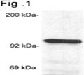
-Western-Blot-NB300-731-img0017.jpg) | Western Blot: GR/NR3C1 Antibody (BuGR2) [NB300-731] - Analysis of glucocorticoid receptor on mouse liver extract. |
-Immunocytochemistry-Immunofluorescence-NB300-731-img0007.jpg) | Immunocytochemistry/Immunofluorescence: GR/NR3C1 Antibody (BuGR2) [NB300-731] - Analysis of Glucocorticoid Receptor using Glucocorticoid Receptor Monoclonal Antibody (BuGR2) shows staining in U251 Cells. Glucocorticoid Receptor (green), F-Actin staining with Phalloidin (red) and nuclei with DAPI (blue) is shown. Cells were grown on chamber slides and fixed with formaldehyde prior to staining. Cells were probed without (control) or with an antibody recognizing Glucocorticoid Receptor at a dilution of 1:100 over night at 4C, washed with PBS and incubated with a DyLight-488 conjugated. |
-Flow-Cytometry-NB300-731-img0018.jpg) | Flow Cytometry: GR/NR3C1 Antibody (BuGR2) [NB300-731] - Using the Alexa Fluor 647 direct conjugate An intracellular stain was performed on HeLa cells with GR/NR3C1 (BuGR2) antibody NB300-731AF647 (blue) and a matched isotype control NB600-986AF647 (orange). Cells were fixed with 4% PFA and then permeablized with 0.1% saponin. Cells were incubated in an antibody dilution of 2 ug/mL for 30 minutes at room temperature. Both antibodies were conjugated to Alexa Fluor 647. |
-Immunocytochemistry-Immunofluorescence-NB300-731-img0014.jpg) | Immunocytochemistry/Immunofluorescence: GR/NR3C1 Antibody (BuGR2) [NB300-731] - Analysis of Glucocorticoid Receptor using Glucocorticoid Receptor Monoclonal Antibody (BuGR2) shows staining in A549 Cells. Glucocorticoid Receptor (green), F-Actin staining with Phalloidin (red) and nuclei with DAPI (blue) is shown. Cells were grown on chamber slides and fixed with formaldehyde prior to staining. Cells were probed without (control) or with an antibody recognizing Glucocorticoid Receptor at a dilution of 1:100 over night at 4C, washed with PBS and incubated with a DyLight-488 conjugated. |
-Immunocytochemistry-Immunofluorescence-NB300-731-img0006.jpg) | Immunocytochemistry/Immunofluorescence: GR/NR3C1 Antibody (BuGR2) [NB300-731] - Analysis of Glucocorticoid Receptor using Glucocorticoid Receptor Monoclonal Antibody (BuGR2) shows staining in Hela Cells. Glucocorticoid Receptor (green), F-Actin staining with Phalloidin (red) and nuclei with DAPI (blue) is shown. Cells were grown on chamber slides and fixed with formaldehyde prior to staining. Cells were probed without (control) or with an antibody recognizing Glucocorticoid Receptor at a dilution of 1:100 over night at 4C, washed with PBS and incubated with a DyLight-488 conjugated. |
-Flow-Cytometry-NB300-731-img0012.jpg) | Flow Cytometry: GR/NR3C1 Antibody (BuGR2) [NB300-731] - Analysis of GR in Jurkat cells compared to an isotype control (blue). |
-Flow-Cytometry-NB300-731-img0015.jpg) | Flow Cytometry: GR/NR3C1 Antibody (BuGR2) [NB300-731] - Analysis of Glucocorticoid Receptor in NIH/3T3 cells compared to an isotype control (blue). |
-Flow-Cytometry-NB300-731-img0016.jpg) | Flow Cytometry: GR/NR3C1 Antibody (BuGR2) [NB300-731] - Analysis of Glucocorticoid Receptor in Hela cells compared to an isotype control (blue). |
-Flow-Cytometry-NB300-731-img0019.jpg) | Flow Cytometry: GR/NR3C1 Antibody (BuGR2) [NB300-731] - An intracellular stain was performed on Jurkat cells with GR/NR3C1 (BuGR2) antibody NB300-731PE (blue) and a matched isotype control (orange). Cells were fixed with 4% PFA and then permeablized with 0.1% saponin. Cells were incubated in an antibody dilution of 5 ug/mL for 30 minutes at room temperature. Both antibodies were conjugated to Phycoerthrin. |
追加しました。
Background
Steroid receptors are ligand-dependent, intracellular proteins that stimulate transcription of specific genes by binding to specific DNA sequences following activation by the appropriate hormone. Glucocorticoids are a family of steroids necessary for the regulation of energy metabolism and the immune and inflammatory responses. These compounds exert their effect through their interaction with the glucocoticoid receptor (GR) and that complex's subsequent association with DNA. All normal mammalian tissues examined to date have been shown to contain glucocorticoid receptor. The corresponding gene for the glucocorticoid receptor is NR3C1.追加しました。
製品情報は掲載時点のものですが、価格表内の価格については随時最新のものに更新されます。お問い合わせいただくタイミングにより製品情報・価格などは変更されている場合があります。
表示価格に、消費税等は含まれていません。一部価格が予告なく変更される場合がありますので、あらかじめご了承下さい。