
抗GFAP抗体 | Anti-GFAP Antibody
掲載日情報:2018/07/09 現在Webページ番号:48663

世界最大級の抗体製品数を取り扱うNovus Biologicals社のGFAPに対する抗体(anti-GFAP | antibody GFAP)です。Novus Biologicals社の抗体は数多くの学術論文で使用実績があります。
※本製品は研究用です。研究用以外には使用できません。

追加しました。
価格
[在庫・価格 :2026年07月14日 12時55分現在]
| 詳細 | 商品名 |
|
文献数 | ||||||||||||||||||||||||||||||||||||||||||||||||||||||||||||||||||||||||||
|---|---|---|---|---|---|---|---|---|---|---|---|---|---|---|---|---|---|---|---|---|---|---|---|---|---|---|---|---|---|---|---|---|---|---|---|---|---|---|---|---|---|---|---|---|---|---|---|---|---|---|---|---|---|---|---|---|---|---|---|---|---|---|---|---|---|---|---|---|---|---|---|---|---|---|---|---|---|
|
Anti-Glial Fibrillary Acidic Protein, Rabbit-Poly <Anti-GFAP> |
|
73 | |||||||||||||||||||||||||||||||||||||||||||||||||||||||||||||||||||||||||||
|
|||||||||||||||||||||||||||||||||||||||||||||||||||||||||||||||||||||||||||||


[在庫・価格 :2026年07月14日 12時55分現在]
Anti-Glial Fibrillary Acidic Protein, Rabbit-Poly <Anti-GFAP>
文献数: 73
- 商品コード:NB300-141
- メーカー:NOV
- 包装:50μl
- 価格:¥110,000
- 在庫:無(未発注)
- 納期:3~4週間 ※※ 表示されている納期は弊社に在庫がなく、取り寄せた場合の目安納期となります。
- 法規制等:
| 説明文 | レビューあり。Simple Western対応抗体。Keywords:FLJ45472|GFAP astrocytes|GFAP immunohistochemistry|GFAP mouse|GFAP rabbit|GFAP stain|glial fibrillary acidic protein クローン:R40 Genbank No: 2670 Protein Accession No: P14136 |
||
|---|---|---|---|
| 法規制等 | |||
| 保存条件 | -20℃ | 法規備考 | |
| 抗原種 | Porcine | 免疫動物 | Rabbit |
| 交差性 | Bovine/Chicken/Equine/Guinea Pig/Human/Mouse/Porcine/Rabbit/Rat | 適用 | IC,IF,IHC,Simple Western,Western Blot |
| 標識 | Unlabeled | 性状 | Serum |
| 吸収処理 | クラス | IgG | |
| クロナリティ | Polyclonal | フォーマット | |
| 掲載カタログ |
|
||
| 製品記事 | 神経マーカー抗体 神経変性疾患研究用抗体 |
||
| 関連記事 | |||
追加しました。
Image
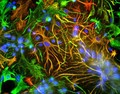
 | Immunocytochemistry/Immunofluorescence: GFAP Antibody [NB300-141] - ICC-IF analysis of mixed neuron-glial cultures using GFAP antibody NB300-141 (red) and Vimentin antibody NB300-223 (green). The fibroblastic cells contain only Vimentin and so are green. The astrocytes contain either Vimentin and GFAP (appearing golden) or predominantly GFAP (appearing red). Blue is nuclear DNA stain. |
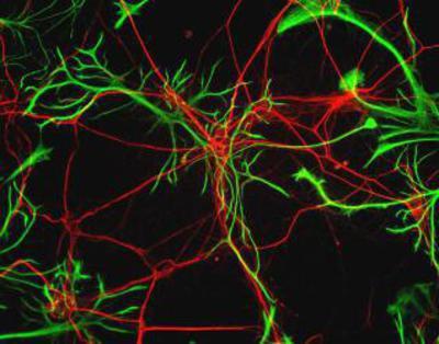 | Immunocytochemistry/Immunofluorescence: GFAP Antibody [NB300-141] - Rat neurons stained with Neurofilament Heavy antibody NB300-217 (red) and GFAP antibody NB300-141 (green). |
 | Immunocytochemistry/Immunofluorescence: GFAP Antibody [NB300-141] - Xenografted mouse brain section : astocyte and human nuclei. Image from a confirmed customer review. |
 | Immunocytochemistry/Immunofluorescence: GFAP Antibody [NB300-141] - Cultured Rat Hippocampal Neuron, image courtesy of customer. |
 | Simple Western: GFAP Antibody [NB300-141] - Simple Western lane view shows a specific band for GFAP in 0.05 mg/ml of Human Brain lysate. This experiment was performed under reducing conditions using the 12-230 kDa separation system. |
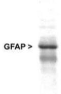
 | Western Blot: GFAP Antibody [NB300-141] - WB analysis of Rat brain lysate using at a dilution of 1:5,000. Specific band running with an apparent SDS-PAGE molecular weight of ~50 kDa corresponds to rodent GFAP was observed. |
 | Western Blot: GFAP Antibody [NB300-141] - Analysis of GFAP expression in whole rat cerebellum homogenate. |
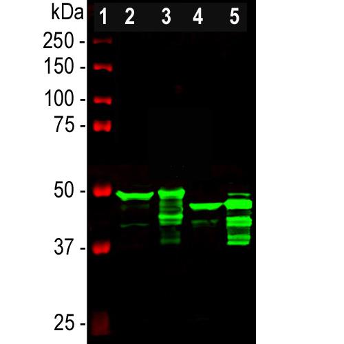 | Western Blot: GFAP Antibody [NB300-141] - Analysis of different tissue lysates using rabbit polyclonal antibody to GFAP, NB300-141, dilution 1:5,000 in green: [1] protein standard (red), [2] rat brain, [3] rat spinal cord, [4] mouse brain, [5] mouse spinal cord. Strong band at about 50kDa corresponds to the major isotype of the GFAP protein. Smaller isotypes and proteolytic fragments of GFAP are also detected on the blot. |
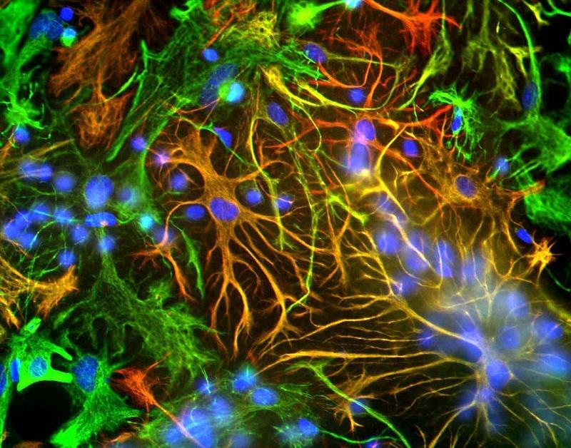
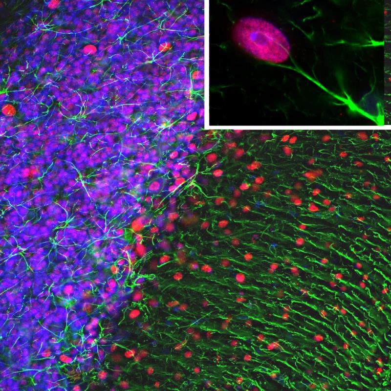 | Immunocytochemistry/Immunofluorescence: GFAP Antibody [NB300-141] - Analysis of a rat cerebellum section stained with rabbit polyclonal antibody to GFAP, NB300-141, dilution 1:5,000 in green and mouse monoclonal antibody to MeCP2, dilution 1:500, in red. The blue is DAPI staining of nuclear DNA. Following transcardial perfusion of rat with 4% paraformaldehyde, brain was post fixed for 1 hour, cut to 45uM, and free-floating sections were stained with above antibodies. The GFAP antibody stains the network of astrocytic cells and the processes of Bergmann glia in the molecular layer. The MeCP2 antibody specifically labels nuclei of certain neurons. |
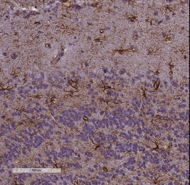 | Immunohistochemistry-Frozen: GFAP Antibody [NB300-141] - Imaging of mouse brain (cortex), 20x magnification. This image was submitted via customer Review. |
追加しました。
Background
GFAP (Glial Fibrillary Acidic Protein) is a member of the class III intermediate filament protein family. It is heavily and specifically expressed in astrocytes and certain other astroglia in the central nervous system, in satellite cells, in peripheral ganglia and in non-myelinating Schwann cells in peripheral nerves. In addition neural stem cells frequently strongly express GFAP. Antibodies to GFAP are therefore very useful as markers of astrocytic cells. In addition many types of brain tumors, presumably derived from astrocytic cells, heavily express GFAP.追加しました。
製品情報は掲載時点のものですが、価格表内の価格については随時最新のものに更新されます。お問い合わせいただくタイミングにより製品情報・価格などは変更されている場合があります。
表示価格に、消費税等は含まれていません。一部価格が予告なく変更される場合がありますので、あらかじめご了承下さい。