
抗Tyrosine Hydroxylase抗体 | Anti-Tyrosine Hydroxylase Antibody
掲載日情報:2018/07/09 現在Webページ番号:48638

世界最大級の抗体製品数を取り扱うNovus Biologicals社のTyrosine Hydroxylaseに対する抗体(anti-Tyrosine Hydroxylase | antibody Tyrosine Hydroxylase)です。Novus Biologicals社の抗体は数多くの学術論文で使用実績があります。
※本製品は研究用です。研究用以外には使用できません。

追加しました。
価格
[在庫・価格 :2026年07月14日 12時55分現在]
| 詳細 | 商品名 |
|
文献数 | ||||||||||||||||||||||||||||||||||||||||||||||||||||||||||||||||||||||||||
|---|---|---|---|---|---|---|---|---|---|---|---|---|---|---|---|---|---|---|---|---|---|---|---|---|---|---|---|---|---|---|---|---|---|---|---|---|---|---|---|---|---|---|---|---|---|---|---|---|---|---|---|---|---|---|---|---|---|---|---|---|---|---|---|---|---|---|---|---|---|---|---|---|---|---|---|---|---|
|
Anti-Tyrosine Hydroxylase, Rabbit-Poly <Anti-TH> |
|
151 | |||||||||||||||||||||||||||||||||||||||||||||||||||||||||||||||||||||||||||
|
|||||||||||||||||||||||||||||||||||||||||||||||||||||||||||||||||||||||||||||
|
Anti-Tyrosine Hydroxylase, Rabbit-Poly |
|
本製品は製品内容等が変更されました | 133 | ||||||||||||||||||||||||||||||||||||||||||||||||||||||||||||||||||||||||||


[在庫・価格 :2026年07月14日 12時55分現在]
Anti-Tyrosine Hydroxylase, Rabbit-Poly <Anti-TH>
文献数: 151
- 商品コード:NB300-109
- メーカー:NOV
- 包装:0.1ml
- 価格:¥122,000
- 在庫:無(未発注)
- 納期:3~4週間 ※※ 表示されている納期は弊社に在庫がなく、取り寄せた場合の目安納期となります。
- 法規制等:
| 説明文 | レビューあり。Simple Western対応抗体。Keywords:anti-TH|DYT14|DYT5b|EC 1.14.16|EC 1.14.16.2|TH|TH mouse|TYH dystonia 14|Tyrosine 3-hydroxylase|tyrosine 3-monooxygenase|tyrosine hydroxylase|Tyrosine Hydroxylase immunohistochemistry|Tyrosine Hydroxylase mouse Genbank No: 7054 Protein Accession No: P07101 |
||
|---|---|---|---|
| 法規制等 | |||
| 保存条件 | -20℃ | 法規備考 | |
| 抗原種 | Rat | 免疫動物 | Rabbit |
| 交差性 | Chicken/Drosophila/Human/Insect/Mammal/Mouse/Rat | 適用 | IC,IF,IHC,Simple Western,Western Blot |
| 標識 | Unlabeled | 性状 | Antigen Affinity Purified |
| 吸収処理 | クラス | IgG | |
| クロナリティ | Polyclonal | フォーマット | |
| 掲載カタログ |
|
||
| 製品記事 | 神経マーカー抗体 神経変性疾患研究用抗体 |
||
| 関連記事 | |||
Anti-Tyrosine Hydroxylase, Rabbit-Poly
文献数: 133
- 商品コード:NB300-109
- メーカー:NOV
- 包装:25μl
- 本製品は製品内容等が変更されました
追加しました。
Image
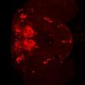
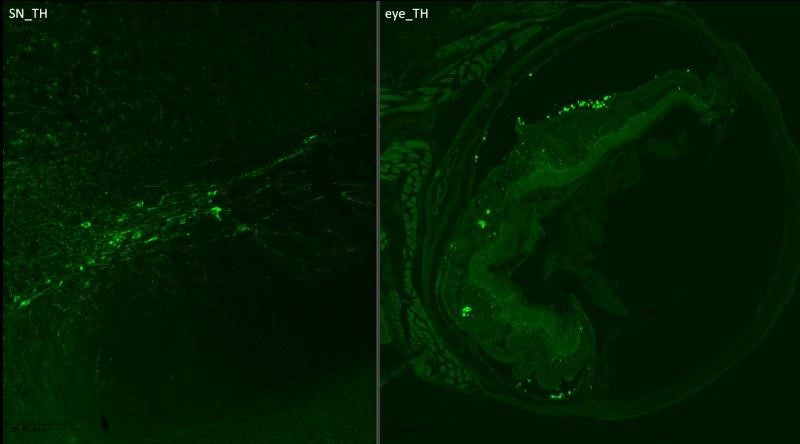
 | Immunocytochemistry/Immunofluorescence: Tyrosine Hydroxylase Antibody [NB300-109] - Dopamine neurons in the mouse substantia nigra. Image from confirmed customer review. |
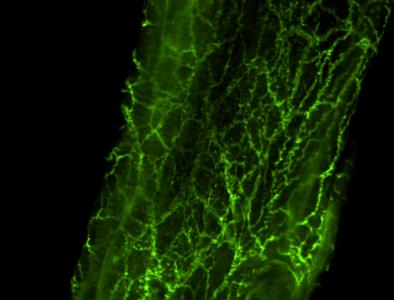 | Immunocytochemistry/Immunofluorescence: Tyrosine Hydroxylase Antibody [NB300-109] - Analysis of Tyrosine Hydroxylase in rat mesenteric artery-whole. Image from verified customer review. |
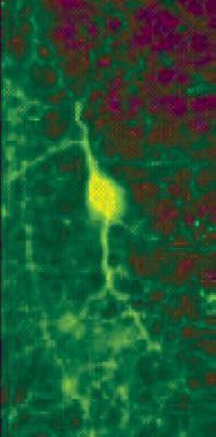 | Immunohistochemistry: Tyrosine Hydroxylase Antibody [NB300-109] - Immunohistochemical staining of retina tissue using TH antibody, NB300-109. |
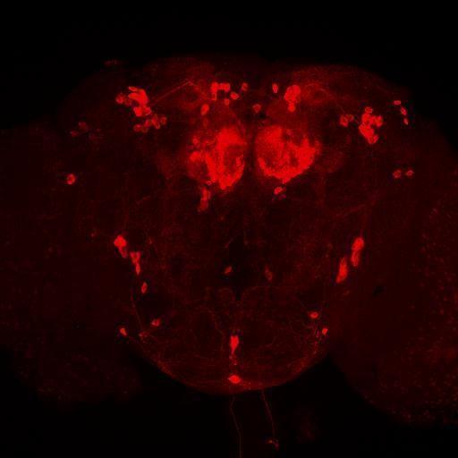 | Immunocytochemistry/Immunofluorescence: Tyrosine Hydroxylase Antibody [NB300-109] - Immunostaining of whole-mount Drosophila brains using NB300-109 at 1:500 dilution. The tyrosine hydroxylase antibody worked really well and produced a bright staining with almost no background. Data courtesy of Dr. Olga Alekseenko, Neurobiology Dept. Harvard Medical School. |
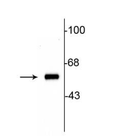 | Western Blot: Tyrosine Hydroxylase Antibody [NB300-109] - Western blot of 10 ug of rat striatal lysate showing specificimmunolabeling of the ~60 kDa tyrosine hydroxylase protein |
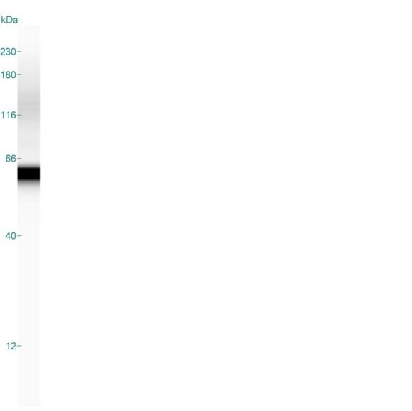 | Simple Western: Tyrosine Hydroxylase Antibody [NB300-109] - Simple Western lane view shows a specific band for Tyrosine Hydroxylase in 0.2 mg/ml of PC-12 lysate. This experiment was performed under reducing conditions using the 12-230 kDa separation system. |
 | Immunocytochemistry: Tyrosine Hydroxylase Antibody [NB300-109] - 10x Magnification cryo-sections mouse: primary antibody: anti-rb TH: 1:500; o.n. secondary antibody: 1:500 dk-anti-rb DyLight488. This image was submitted via customer Review. |
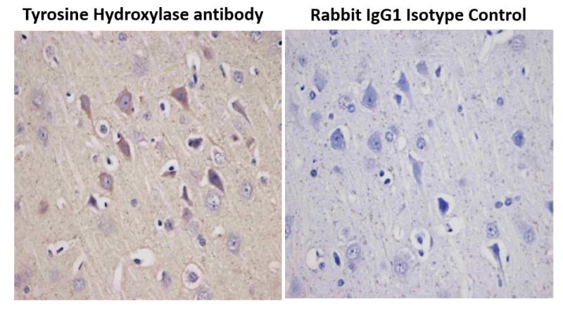 | Immunohistochemistry-Paraffin: Tyrosine Hydroxylase Antibody [NB300-109] - Immunohistochemical analysis of a formalin fixed and paraffin embedded rat brain tissue section using Tyrosine Hydroxylase antibody at 1:5000 dilution. The primary antibody binding to its antigen was detected using HRP anti-Polyvalent ready-to-use kit (Ultratek, 35368) with DAB (brown) and the sections were further counterstained using hematoxylin. Isotype control section was incubated with Rabbit IgG Isotype Control antibody and was processed under the same assay conditions. Tyrosine hydroxylase staining (brown) was observed in the test samples only and the signal was specifically localized to the neuronal cells. |
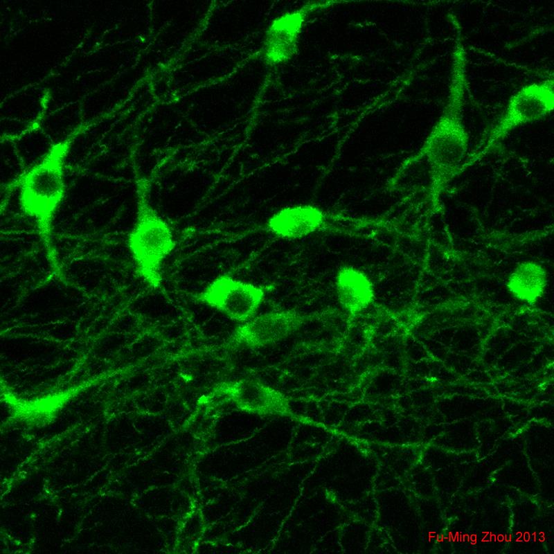
 | Immunocytochemistry/Immunofluorescence: Tyrosine Hydroxylase Antibody [NB300-109] - Rat midbrain mixed neuronal cultures showing TH positive neurons in green and MAP2 in red. Image courtesy of Aurelie de Rus Jacquet,laboratory of Dr. Jean-Christophe Rochet, Purdue University. |
追加しました。
Background
Tyrosine hydroxylase (TH) is the rate-limiting enzyme in the synthesis of the catecholamines dopamine, epinephrine and norepinephrine. Therefore the regulation of the TH enzyme represents the central means for controlling the synthesis of these important catecholamines. TH has a large molecular diversity, resulting from differential splicing of its mRNA, which is tissue specific and might result in long term changes in activity of the enzyme and its availability of neurotransmitter at various synapses. The presence of different DNA sequences at the TH locus confers susceptibility to various disorders of the brain including manic-depression and schizophrenia. Parkinson's disease is also considered a TH deficiency as low dopamine levels are a consistent neurochemical abnormality.追加しました。
製品情報は掲載時点のものですが、価格表内の価格については随時最新のものに更新されます。お問い合わせいただくタイミングにより製品情報・価格などは変更されている場合があります。
表示価格に、消費税等は含まれていません。一部価格が予告なく変更される場合がありますので、あらかじめご了承下さい。