
抗Ki-67/MKI67抗体 | Anti-Ki-67/MKI67 Antibody
掲載日情報:2018/07/09 現在Webページ番号:48363

世界最大級の抗体製品数を取り扱うNovus Biologicals社のKi-67/MKI67に対する抗体(anti-Ki-67/MKI67 | antibody Ki-67/MKI67)です。Novus Biologicals社の抗体は数多くの学術論文で使用実績があります。
※本製品は研究用です。研究用以外には使用できません。
価格表左の「文献」アイコンから、使用文献情報一覧が表示できます。

カートに商品を
追加しました。
追加しました。
価格
[在庫・価格 :2026年07月22日 08時55分現在]
※ 表示されている納期は弊社に在庫が無く、取り寄せた場合の納期目安となります。
| 詳細 | 商品名 |
|
文献数 | ||||||||||||||||||||||||||||||||||||||||||||||||||||||||||||||||||||||||||||||||||
|---|---|---|---|---|---|---|---|---|---|---|---|---|---|---|---|---|---|---|---|---|---|---|---|---|---|---|---|---|---|---|---|---|---|---|---|---|---|---|---|---|---|---|---|---|---|---|---|---|---|---|---|---|---|---|---|---|---|---|---|---|---|---|---|---|---|---|---|---|---|---|---|---|---|---|---|---|---|---|---|---|---|---|---|---|---|
|
Anti-Ki67, Rabbit-Poly |
|
63 | |||||||||||||||||||||||||||||||||||||||||||||||||||||||||||||||||||||||||||||||||||
|
|||||||||||||||||||||||||||||||||||||||||||||||||||||||||||||||||||||||||||||||||||||


[在庫・価格 :2026年07月22日 08時55分現在]
※ 表示されている納期は弊社に在庫が無く、取り寄せた場合の納期目安となります。
Anti-Ki67, Rabbit-Poly
文献数: 63
- 商品コード:NB110-89717
- メーカー:NOV
- 包装:0.1ml
- 価格:¥108,000
- 在庫:無(未発注)
- 納期:3~4週間 ※※ 表示されている納期は弊社に在庫がなく、取り寄せた場合の目安納期となります。
- 法規制等:
| 説明文 | レビューあり。Keywords:antigen identified by monoclonal Ki-67|antigen KI-67|Ki67|Ki-67|KIA|Marker Of Proliferation Ki-67|MIB-1|PPP1R105|proliferation-related Ki-67 antigen|Protein Phosphatase 1|Regulatory Subunit 105 Genbank No: 4288 |
||||||
|---|---|---|---|---|---|---|---|
| 別包装品 | 別包装品あり | ||||||
| 法規制等 | |||||||
| 保存条件 | -20℃ | 法規備考 | |||||
| 抗原種 | Mouse | 免疫動物 | Rabbit | ||||
| 交差性 | Human/Mouse/Porcine/Rat | 適用 | FCM,IC,IF,IHC,Western Blot | ||||
| 標識 | Unlabeled | 性状 | Antigen Affinity Purified | ||||
| 吸収処理 | クラス | IgG | |||||
| クロナリティ | Polyclonal | フォーマット | |||||
| 掲載カタログ |
|
||||||
| 製品記事 | 抗Ki67抗体(Anti-Ki67 Antibody) 抗Ki-67/MKI67抗体 | Anti- Ki-67/MKI67 antibody |
||||||
| 関連記事 | |||||||
カートに商品を
追加しました。
追加しました。
Image
 | Immunohistochemistry-Paraffin: Ki-67/MKI67 Antibody [NB110-89717] - Staining of a cross section of mouse spleen. Detection: DAB staining using Immunohistochemistry Accessory Kit. Epitope Retrieval Buffer-High pH was substituted for Epitope Retrieval Buffer-Reduced pH. |
 | Immunohistochemistry: Ki-67/MKI67 Antibody [NB110-89717] - Ki67 Antibody [NB110-89717] - FFPE section of mouse peyer's patch. Antibody: Affinity purified rabbit anti-mouse Ki-67 used at a dilution of 1:250. Detection: DAB staining using Immunohistochemistry Accessory Kit. Epitope Retrieval Buffer-High pH was substituted for Epitope Retrieval Buffer-Reduced pH. |
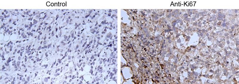
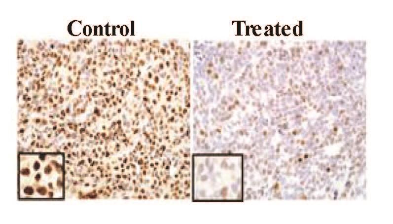 | Immunohistochemistry-Paraffin: Ki-67/MKI67 Antibody [NB110-89717] - analysis of Ki-67 in human prostate xenograft control (left) and treated (right) using anti-Ki-67 antibody. Image from verified customer review. |
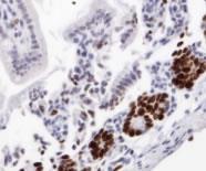
 | Immunohistochemistry-Paraffin: Ki-67/MKI67 Antibody [NB110-89717] - analysis of Ki-67 in paraffin embedded mouse prostate tissue using anti-Ki-67 antibody. Image from verified customer review. |
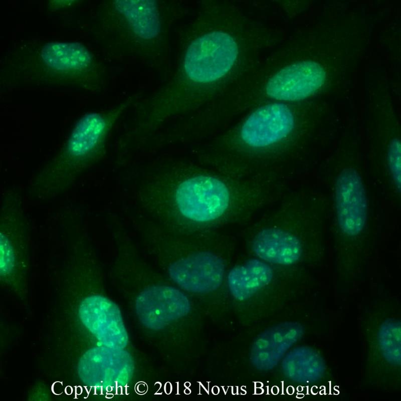 | Immunocytochemistry/Immunofluorescence: Ki-67/MKI67 Antibody [NB110-89717] - HeLa cells were fixed for 10 minutes using 10% formalin and then permeabilized for 5 minutes using 1X PBS + 0.5% Triton-X100. The cells were incubated with anti-Ki-67/MKI67 at 2 ug/ml overnight at 4C and detected with an anti-rabbit Dylight 488 (Green) at a 1:500 dilution. Nuclei were counterstained with DAPI (Blue). Cells were imaged using a 40X objective. |
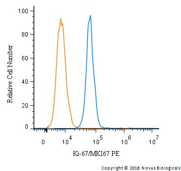 | Flow Cytometry: Ki-67/MKI67 Antibody [NB110-89717] - An intracellular stain was performed on U-937 cells with Ki-67/MKI67 Antibody NB110-89717PE (blue) and a matched isotype control (orange). Cells were fixed with 4% PFA and then permeabilized with 0.1% saponin. Cells were incubated in an antibody dilution of 2.5 ug/mL for 30 minutes at room temperature. Both antibodies were conjugated to Phycoerythrin. |
 | Immunohistochemistry: Ki67 Antibody [NB110-89717] - Detection of Ki67 in formalin-fixed paraffin embedded mouse intestine using NB110-89717. |
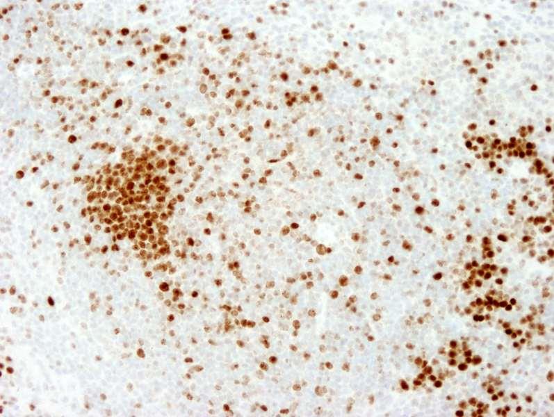
 | Immunohistochemistry: Ki67 Antibody [NB110-89717] - Immunohistochemical analysis of mouse spleen. |
 | Immunohistochemistry: Ki-67/MKI67 Antibody [NB110-89717] - Analysis of a mouse intestine cross section. The antibody was used at a dilution of 1:250. Detection: DAB staining. Epitope Retrieval Buffer-High pH was substituted for Epitope Retrieval Buffer-Reduced pH. |
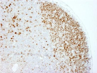
 | Immunohistochemistry-Paraffin: Ki-67/MKI67 Antibody [NB110-89717] - Analysis of Ki-67 in a formalin fixed paraffin embedded cross section of mouse intestine. Detection: DAB staining using Immunohistochemistry Accessory Kit. Epitope Retrieval Buffer-High pH was substituted for Epitope Retrieval Buffer-Reduced pH. |
 | Flow Cytometry: Ki67 Antibody [NB110-89717] - Staining of mouse bone marrow cells using NB110-89717 at a dilution of 1:100. Photo courtesy of product review by verified customer. |
カートに商品を
追加しました。
追加しました。
Background
Originally discovered employing mouse monoclonal antibody against a nuclear antigen from Hodgkin's lymphoma-derived cell line, this non-histone protein was named Ki67 after researcher's location (Gerdes and colleagues), Ki for Kiel University in Germany and 67 referring to the clone number on the 96-well plate. It interacts with KIF15 as well as MKI67IP, and is suggested to be involved in cell cycle regulation. Ki67 is a large protein with expected molecular weight of about 395 kD and has a very complex localization pattern within the nucleus, one which changes during cell cycle progression. Its expression occurs specially during late G1, S, G2 and M phases of the cell cycle, while in cells undergoing G0 phase, Ki67 remains undetectable. Ki67 undergoes phosphorylation/dephosphorylation during mitosis, is susceptible to proteases and its structure implies that its expression is regulated by proteolytic pathways. Ki67 is associated with nucleolar DFC (dense fibrillary component) and its regulation appears to be tightly controlled (estimated half life is 60-90 min, regardless of the cell position in the cell cycle), presumably by precise synthesis and degradation systems involving proteasome, a protease complex. Due to its association with cell divison process, Ki-67 is routinely used as cellular proliferation marker of solid tumors as well as certain hematological malignancies, and a correlation has been demonstrated between Ki-67 index and the histopathological grade of cancers.カートに商品を
追加しました。
追加しました。
製品情報は掲載時点のものですが、価格表内の価格については随時最新のものに更新されます。お問い合わせいただくタイミングにより製品情報・価格などは変更されている場合があります。
表示価格に、消費税等は含まれていません。一部価格が予告なく変更される場合がありますので、あらかじめご了承下さい。