抗Beclin 1抗体 | Anti-Beclin 1 Antibody
掲載日情報:2018/07/09 現在Webページ番号:48356
世界最大級の抗体製品数を取り扱うNovus Biologicals社のBeclin 1に対する抗体(anti-Beclin 1 | antibody Beclin 1)です。Novus Biologicals社の抗体は数多くの学術論文で使用実績があります。
※本製品は研究用です。研究用以外には使用できません。
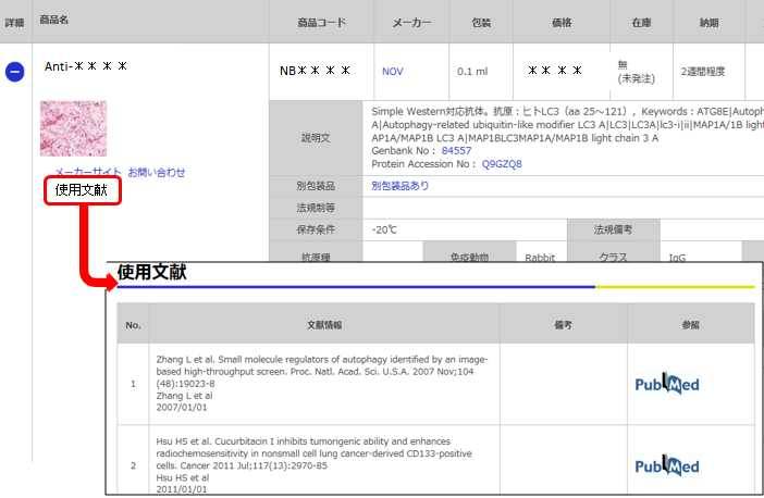
価格表左の「文献」アイコンから、使用文献情報一覧が表示できます。


カートに商品を
追加しました。
追加しました。
価格
[在庫・価格 :2026年02月23日 16時35分現在]
※ 表示されている納期は弊社に在庫が無く、取り寄せた場合の納期目安となります。
| 詳細 | 商品名 |
|
文献数 | ||||||||||||||||||||||||||||||||||||||||||||||||||||||||||||||||||||||||||||||||||
|---|---|---|---|---|---|---|---|---|---|---|---|---|---|---|---|---|---|---|---|---|---|---|---|---|---|---|---|---|---|---|---|---|---|---|---|---|---|---|---|---|---|---|---|---|---|---|---|---|---|---|---|---|---|---|---|---|---|---|---|---|---|---|---|---|---|---|---|---|---|---|---|---|---|---|---|---|---|---|---|---|---|---|---|---|---|
|
Anti-Beclin1, Rabbit-Poly |
|
48 | |||||||||||||||||||||||||||||||||||||||||||||||||||||||||||||||||||||||||||||||||||
|
|||||||||||||||||||||||||||||||||||||||||||||||||||||||||||||||||||||||||||||||||||||
[在庫・価格 :2026年02月23日 16時35分現在]
※ 表示されている納期は弊社に在庫が無く、取り寄せた場合の納期目安となります。
Anti-Beclin1, Rabbit-Poly
文献数: 48
- 商品コード:NB110-87318
- メーカー:NOV
- 包装:0.05ml
- 価格:¥96,000
- 在庫:無(未発注)
- 納期:3~4週間 ※※ 表示されている納期は弊社に在庫がなく、取り寄せた場合の目安納期となります。
- 法規制等:
| 説明文 | レビューあり。Simple Western対応抗体。Keywords:ATG6|ATG6 autophagy related 6 homolog|beclin 1 (coiled-coil|moesin-like BCL2 interacting protein)|moesin-like BCL2-interacting protein)|beclin 1|autophagy related|beclin1|beclin-1|Coiled-coil myosin-like BCL2-interacting protein|GT197|Protein GT197 Genbank No: 8678 Protein Accession No: Q14457 |
||||||
|---|---|---|---|---|---|---|---|
| 別包装品 | 別包装品あり | ||||||
| 法規制等 | |||||||
| 保存条件 | -20℃ | 法規備考 | |||||
| 抗原種 | 免疫動物 | Rabbit | |||||
| 交差性 | Alligator/Canine/Human/Mouse/Porcine/Primate/Rat | 適用 | IC,IF,IHC,Simple Western,Western Blot | ||||
| 標識 | Unlabeled | 性状 | Antigen Affinity Purified | ||||
| 吸収処理 | クラス | IgG | |||||
| クロナリティ | Polyclonal | フォーマット | |||||
| 掲載カタログ |
|
||||||
| 製品記事 | 抗Beclin 1抗体 | Anti- Beclin 1 antibody 抗 Beclin 1 抗体 |
||||||
| 関連記事 | |||||||
カートに商品を
追加しました。
追加しました。
Image
カートに商品を
追加しました。
追加しました。
Background
Beclin 1 is the first identified mammalian gene to mediate autophagy and also has tumor suppressor and antiviral function. Autophagy, a process of bulk protein degradation through an autophagosomic-lysosomal pathway, is important for differentiation, survival during nutrient deprivation, and normal growth control, and is often defective in tumor cells. Beclin 1 was originally isolated in a yeast two hybrid screen to identify Bcl-2-binding partners and maps to a tumor susceptibility locus on human chromosome 17q21 that is frequently monoallelically deleted in human breast, ovarian and prostate cancer. Beclin 1 encodes an evolutionarily conserved 60 kDa coiled coil protein that is expressed in human muscle, epithelial cells and neurons. In gene transfer studies, Beclin 1 promotes nutrient deprivation-induced autophagy, inhibits mammary tumorigenesis, and inhibits viral replication. Expression of the Beclin 1 protein is frequently decreased in malignant breast epithelial cells. Based upon these observations, it is speculated that beclin 1 may work through induction of autophagy to negatively regulate tumorigenesis and to control viral infections. Beclin 1 may also play a role in other biological processes in which autophagy is important such as cell differentiation and nutritional stress responses.カートに商品を
追加しました。
追加しました。
製品情報は掲載時点のものですが、価格表内の価格については随時最新のものに更新されます。お問い合わせいただくタイミングにより製品情報・価格などは変更されている場合があります。
表示価格に、消費税等は含まれていません。一部価格が予告なく変更される場合がありますので、あらかじめご了承下さい。


