
抗HIF-1 alpha Pack抗体 | Anti-HIF-1 alpha Pack Antibody
掲載日情報:2018/07/09 現在Webページ番号:47787

世界最大級の抗体製品数を取り扱うNovus Biologicals社のHIF-1 alpha Packに対する抗体(anti-HIF-1 alpha Pack | antibody HIF-1 alpha Pack)です。Novus Biologicals社の抗体は数多くの学術論文で使用実績があります。
※本製品は研究用です。研究用以外には使用できません。

追加しました。
価格
[在庫・価格 :2026年07月17日 00時01分現在]
| 詳細 | 商品名 |
|
文献数 | ||||||||||||||||||||||||||||||||||||||||||||||||||||||||||||||||||||||||||
|---|---|---|---|---|---|---|---|---|---|---|---|---|---|---|---|---|---|---|---|---|---|---|---|---|---|---|---|---|---|---|---|---|---|---|---|---|---|---|---|---|---|---|---|---|---|---|---|---|---|---|---|---|---|---|---|---|---|---|---|---|---|---|---|---|---|---|---|---|---|---|---|---|---|---|---|---|---|
|
Anti-HIF-1α Immunohistochemistry Antibody Sample Pack |
|
本製品は取扱中止になりました | 6 | ||||||||||||||||||||||||||||||||||||||||||||||||||||||||||||||||||||||||||
|
|||||||||||||||||||||||||||||||||||||||||||||||||||||||||||||||||||||||||||||


[在庫・価格 :2026年07月17日 00時01分現在]
Anti-HIF-1α Immunohistochemistry Antibody Sample Pack
文献数: 6
- 商品コード:NB100-901IHC
- メーカー:NOV
- 包装:1set
- 本製品は取扱中止になりました
| 説明文 | Genbank No:
3091
|
||
|---|---|---|---|
| 法規制等 | |||
| 保存条件 | 法規備考 | ||
| 抗原種 | Human | 免疫動物 | |
| 交差性 | Human/Mouse/Rat | 適用 | FCM,IC,IF,IHC,IP,Simple Western,Western Blot |
| 標識 | Unlabeled | 性状 | |
| 吸収処理 | クラス | ||
| クロナリティ | フォーマット | ||
| 掲載カタログ |
|
||
| 製品記事 | 抗HIF抗体(Anti-HIF-1/HIF-2 antibody) |
||
| 関連記事 | |||
追加しました。
Image
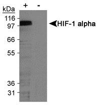
 | Western Blot: HIF-1 alpha Antibody Pack [NB100-901IHC] - Detection of mouse HIF1-alpha on hypoxia treated MEFs using NB 100-449. |
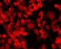
 | Immunocytochemistry/Immunofluorescence: HIF-1 alpha Antibody Pack [NB100-901IHC] - Detection of HIF-1 alpha (red dye 568) in a cultured raw mouse macrophage cell line, using NB100-131. 100X magnification. Photos courtesy of Susan Alexander and Hattie Gresham, PhD. |
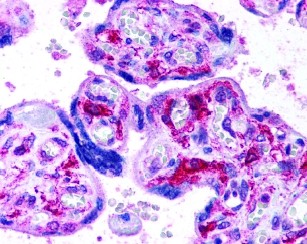 | Immunohistochemistry: HIF-1 alpha Antibody Pack [NB100-901IHC] - Staining of placenta, villi using. |
 | Flow Cytometry: HIF-1 alpha Antibody Pack [NB100-901IHC] - Analysis of HIF1-alpha in CoCl2 Treated vs. Untreated HeLa Cells. Hela cells were treated for 15 hrs with 200uM CoCl2, fixed in PFA, and permeabilized in 90% MeOH. One million cells were stained with 0.125ug of NB100-449 and secondary FITC-conjugated goat anti-rabbit (in a 150ul reaction). Black- treated, anti-KLH control IgG; Reduntreated, anti-HIF1-alpha; Blue- treated, anti-HIF1-alpha. |
 | Western Blot: HIF-1 alpha Antibody Pack [NB100-901IHC] - Detection of HIF-1 alpha in cobalt chloride treated/untreated COS-7 nuclear extracts. |
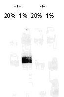 | Western Blot: HIF-1 alpha Antibody Pack [NB100-901IHC] - On day 1, MEF cells (+/+,-/-), were grown on 15cm dish (2x10 to the 6th cells). On day 2, cells were exposed to hypoxia for 4hrs. Cells were washed with ice cold PBS twice and whole cell protein was extracted with RIPA buffer fortified with protease. Upon quantification, 100ug of protein was fractionated on 7% polyacralymide gel. -Gel was transferred overnight onto nitrocellulose membrane. The membrane was probed with HIF-1 alpha monoclonal antibody at a 1:500 dilution (NB 100-123). The secondary antibody was conjugated with HRP and was used at a 1:2500 dilution. Photo courtesy of Dr. Gregg Semenza, Johns Hopkins University. |
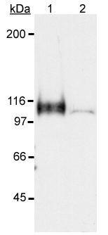 | Western Blot: HIF-1 alpha Antibody Pack [NB100-901IHC] - Detection of HIF-1 alpha in a hypoxic sample. Lane 1: CoCl2 treated Cos-7 nuclear extract (hypoxic). Lane 2: Untreated Cos-7 nuclear extract (normoxic) NB100-449. |
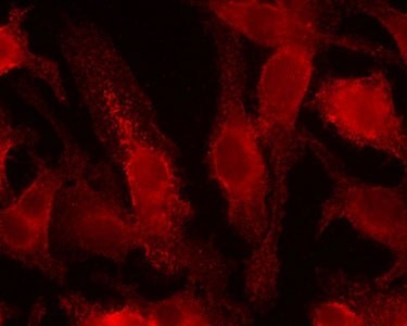
 | Immunocytochemistry/Immunofluorescence: HIF-1 alpha Antibody Pack [NB100-901IHC] - Detection of HIF-1 alpha (red dye) in a cell cytospin from a lavage of a murine skin pouch infected with Staph Aureus, using NB100-131. Blue dye is DAPI nuclear staining. Photos courtesy of Susan Alexander and Hattie Gresham, PhD. |
 | Immunocytochemistry/Immunofluorescence: HIF-1 alpha Antibody Pack [NB100-901IHC] - Detection of HIF-1 alpha (red dye) in a cell cytospin from a lavage of a murine skin pouch infected with Staph Aureus, using NB100-131. 100X magnification. Blue dye is DAPI nuclear staining. Photos courtesy of Susan Alexander and Hattie Gresham, PhD. |
 | Immunocytochemistry/Immunofluorescence: HIF-1 alpha Antibody Pack [NB100-901IHC] - Detection of HIF-1 alpha (red dye 568) in a cultured raw mouse macrophage cell line. Photos courtesy of Susan Alexander and Hattie Gresham, PhD. |
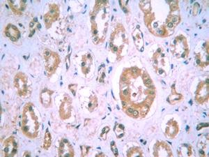 | Immunohistochemistry: HIF-1 alpha Antibody Pack [NB100-901IHC] - Staining of HIF-1 alpha in human kidney. |
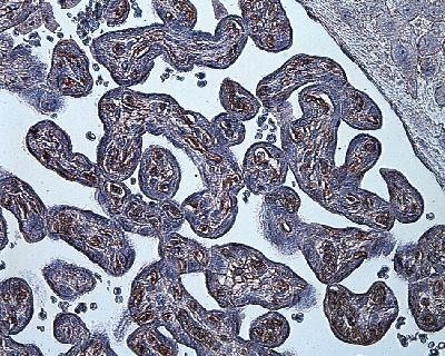 | Immunohistochemistry: HIF-1 alpha Antibody Pack [NB100-901IHC] - Staining in hypoxia-induced human placenta. |
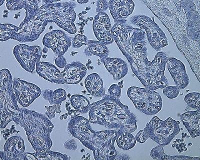 | Immunohistochemistry-Paraffin: HIF-1 alpha Antibody Pack [NB100-901IHC] - Negative control stain of human placenta (from sea level) using mouse IgG at 1:100. 4uM paraffin-embedded section. |
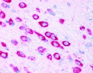 | Immunohistochemistry: HIF-1 alpha Antibody Pack [NB100-901IHC] - Staining of mouse brain, neurons using NB100-449. |
 | Immunoprecipitation: HIF-1 alpha Antibody Pack [NB100-901IHC] - Immunoprecipitation of HIF-1alpha using NB100-123. Heavy and light chains are also detected. |
追加しました。
Background
Our HIF-1 alpha sample pack provides a convenient way to determine the optimal HIF-1 alpha antibody for your specific species and tissues. Hypoxia contributes significantly to the pathophysiology of major categories of human disease, including myocardial and cerebral ischemia, cancer, pulmonary hypertension, congenital heart disease and chronic obstructive pulmonary disease. HIF-1 is a nuclear protein that activates gene transcription in response to reduced cellular 02 concentration. HIF-1 activates the transcription of EPO, VEGF, iNOS, heme oxygenase 1 and several other critical intracellular responses to hypoxia. HIF-1 is a heterodimer composed of HIF-1 alpha and HIF-1 beta subunits. Both subunits are induced by hypoxia and rapidly decay upon return to normoxia and are basic-helix-loop-helix-PAS proteins. Recent research indicates the ability to regulate hypoxia-inducible factors may be related to tumor-related angiogenesis in certain cancers.追加しました。
製品情報は掲載時点のものですが、価格表内の価格については随時最新のものに更新されます。お問い合わせいただくタイミングにより製品情報・価格などは変更されている場合があります。
表示価格に、消費税等は含まれていません。一部価格が予告なく変更される場合がありますので、あらかじめご了承下さい。