
抗RelA/NFkB p65 (112A1021)抗体 | Anti-RelA/NFkB p65 (112A1021) Antibody
掲載日情報:2018/07/09 現在Webページ番号:47260

世界最大級の抗体製品数を取り扱うNovus Biologicals社のRelA/NFkB p65 (112A1021)に対する抗体(anti-RelA/NFkB p65 (112A1021) | antibody RelA/NFkB p65 (112A1021))です。Novus Biologicals社の抗体は数多くの学術論文で使用実績があります。
※本製品は研究用です。研究用以外には使用できません。

追加しました。
価格
[在庫・価格 :2026年07月14日 12時55分現在]
| 詳細 | 商品名 |
|
文献数 | ||||||||||||||||||||||||||||||||||||||||||||||||||||||||||||||||||||||||||||||||||
|---|---|---|---|---|---|---|---|---|---|---|---|---|---|---|---|---|---|---|---|---|---|---|---|---|---|---|---|---|---|---|---|---|---|---|---|---|---|---|---|---|---|---|---|---|---|---|---|---|---|---|---|---|---|---|---|---|---|---|---|---|---|---|---|---|---|---|---|---|---|---|---|---|---|---|---|---|---|---|---|---|---|---|---|---|---|
|
Anti-RelA/NFkB p65, Mouse-Mono(112A1021) |
|
13 | |||||||||||||||||||||||||||||||||||||||||||||||||||||||||||||||||||||||||||||||||||
|
|||||||||||||||||||||||||||||||||||||||||||||||||||||||||||||||||||||||||||||||||||||


[在庫・価格 :2026年07月14日 12時55分現在]
Anti-RelA/NFkB p65, Mouse-Mono(112A1021)
文献数: 13
- 商品コード:NB100-56712
- メーカー:NOV
- 包装:0.1mg
- 価格:¥110,000
- 在庫:無(未発注)
- 納期:3~4週間 ※※ 表示されている納期は弊社に在庫がなく、取り寄せた場合の目安納期となります。
- 法規制等:
| 説明文 | レビューあり。Simple Western対応抗体。旧IMGENEX社 商品コード:IMG-150A,Keywords:anti-NF kB p65|NFkB p65|NF-kB p65|p65|RelA|rela p65|MGC131774|nf kb p65|NFKB3v-rel avian reticuloendotheliosis viral oncogene homolog A (nuclear factor ofkappa light polypeptide gene enhancer in B-cells 3 (p65))|Nuclear factor NF-kappa-B p65 subunit クローン:112A1021 Genbank No: 5970 Protein Accession No: Q969V5 |
||||||
|---|---|---|---|---|---|---|---|
| 別包装品 | 別包装品あり | ||||||
| 法規制等 | |||||||
| 保存条件 | -20℃ | 法規備考 | |||||
| 抗原種 | 免疫動物 | Mouse | |||||
| 交差性 | Human/Mouse/Rat | 適用 | FCM,IHC,IP,Simple Western,Western Blot | ||||
| 標識 | Unlabeled | 性状 | Protein A/G Affinity Purified | ||||
| 吸収処理 | クラス | IgG | |||||
| クロナリティ | Monoclonal | フォーマット | |||||
| 掲載カタログ |
|
||||||
| 製品記事 | 神経変性疾患研究用抗体 |
||||||
| 関連記事 | |||||||
追加しました。
Image
-Western-Blot-NB100-56712-img0016.jpg) | Western Blot: RelA/NFkB p65 Antibody (112A1021) [NB100-56712] - Lysates of HeLa human cervical epithelial carcinoma cell line and Daudi human Burkitt's lymphoma cell line. PVDF membrane was probed with 1 ug/mL mouse anti-RelA/NFkB p65 monoclonal (NB100-56712, Novus Biologicals), followed by 1:2000 dilution of the appropriate HRP-conjugated secondary antibody, donkey anti-mouse IgG (HAF018). |
-Immunohistochemistry-Paraffin-NB100-56712-img0011.jpg) | Immunohistochemistry-Paraffin: RelA/NFkB p65 Antibody (112A1021) [NB100-56712] - Analysis using Azide Free version of NB100-56712. Formalin-fixed, paraffin-embedded ovarian cystadenocarcinoma probed with p65 antibody at 5 ug/ml. Human tissue TMA was used for this test. Staining of formalin-fixed tissues is enhanced by boiling tissue s |
-Flow-(Intracellular)-NB100-56712-img0012.jpg) | Flow (Intracellular): RelA/NFkB p65 Antibody (112A1021) [NB100-56712] - Analysis using Azide Free version of NB100-56712. Intracellular staining of 293 HEK cells using 0.5 ug of p65 antibody. Green histogram represents the isotype control (p65) antibody. this antibody was used for this test, and an anti-mouse IgG PE conjugate |
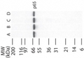
-Western-Blot-NB100-56712-img0007.jpg) | Western Blot: RelA/NFkB p65 Antibody (112A1021) [NB100-56712] - Analysis of NF-kB (p65) using NB100-56055 (biotinylated version of Clone 112A1021) at 2 ug/ml in 30 ug of A) Ramos, B) Daudi, C) HeLa and D) mouse NIH 3T3 cell lysate. |
-Western-Blot-NB100-56712-img0010.jpg) | Western Blot: RelA/NFkB p65 Antibody (112A1021) [NB100-56712] - Analysis of Biotin conjugate of NB100-56712. OfRelA/NFkB p65 Antibody (112A1021) [Biotin]using NB100-56055 at 2 ug/ml in 30 ug of A) Ramos, B) Daudi, C) HeLa and D) mouse NIH 3T3 cell lysate. |
-Western-Blot-NB100-56712-img0013.jpg) | Western Blot: RelA/NFkB p65 Antibody (112A1021) [NB100-56712] - Analysis using Azide Free version of NB100-56712. P65 using p65 antibody at 2 ug/ml in 30 ug of A) Ramos, B) Daudi, C) HeLa and D) mouse NIH 3T3 cell lysate. |
-Immunohistochemistry-Paraffin-NB100-56712-img0009.jpg) | Immunohistochemistry-Paraffin: RelA/NFkB p65 Antibody (112A1021) [NB100-56712] - Formalin-fixed, paraffin-embedded ovarian cystadenocarcinoma probed with p65 antibody at 5 ug/ml. Human tissue TMA was used for this test. Staining of formalin-fixed tissues is enhanced by boiling tissue sections in 10 mM sodium citrate buffer, pH 6.0 for 10-20 min followed by cooling at RT for 20 min. |
-Flow-Cytometry-NB100-56712-img0004.jpg) | Flow Cytometry: RelA/NFkB p65 Antibody (112A1021) [NB100-56712] - Intracellular staining of 293 HEK cells using 0.5 ug of p65 antibody. Green histogram represents the isotype control (p65) antibody. This NFkB p65 antibody was used for this test with an anti-mouse IgG PE conjugated secondary antibody. |
-Simple-Western-NB100-56712-img0006.jpg) | Simple Western: RelA/NFkB p65 Antibody (112A1021) [NB100-56712] - Simple Western lane view shows a specific band for NFkB p65 in 1.0 mg/ml of HeLa lysate. This experiment was performed under reducing conditions using the 12-230 kDa separation system. |
-Simple-Western-NB100-56712-img0014.jpg) | Simple Western: RelA/NFkB p65 Antibody (112A1021) [NB100-56712] - Analysis using Azide Free version of NB100-56712. Simple Western lane view shows a specific band for NFkB p65 in 1.0 mg/ml of HeLa lysate. This experiment was performed under reducing conditions using the 12-230 kDa separation system. |
追加しました。
Background
NF-kB (nuclear factor kB) regulates the expression of a large number of genes that play critical roles in apoptosis, viral replication, tumorigenesis, various autoimmune diseases and inflammation. The active nuclear form of the NF-kB transcription factor complex is composed of two DNA binding subunits, NF-kB p65 and p50, both of which share extensive N-terminal sequence homology with the v-rel oncogene product. N-terminal regions of p50 and p65 are critical for DNA binding and help determine the DNA-binding specificities of p50 and p65.追加しました。
製品情報は掲載時点のものですが、価格表内の価格については随時最新のものに更新されます。お問い合わせいただくタイミングにより製品情報・価格などは変更されている場合があります。
表示価格に、消費税等は含まれていません。一部価格が予告なく変更される場合がありますので、あらかじめご了承下さい。