
抗Caspase-3 (31A1067) - (Pro and Active)抗体 | Anti-Caspase-3 (31A1067) - (Pro and Active) Antibody
掲載日情報:2018/07/09 現在Webページ番号:47258

世界最大級の抗体製品数を取り扱うNovus Biologicals社のCaspase-3 (31A1067) - (Pro and Active)に対する抗体(anti-Caspase-3 (31A1067) - (Pro and Active) | antibody Caspase-3 (31A1067) - (Pro and Active))です。Novus Biologicals社の抗体は数多くの学術論文で使用実績があります。
※本製品は研究用です。研究用以外には使用できません。

追加しました。
価格
[在庫・価格 :2026年07月14日 11時35分現在]
| 詳細 | 商品名 |
|
文献数 | ||||||||||||||||||||||||||||||||||||||||||||||||||||||||||||||||||||||||||||||||||
|---|---|---|---|---|---|---|---|---|---|---|---|---|---|---|---|---|---|---|---|---|---|---|---|---|---|---|---|---|---|---|---|---|---|---|---|---|---|---|---|---|---|---|---|---|---|---|---|---|---|---|---|---|---|---|---|---|---|---|---|---|---|---|---|---|---|---|---|---|---|---|---|---|---|---|---|---|---|---|---|---|---|---|---|---|---|
|
Anti-Caspase-3 (Pro and Active), Mouse-Mono(31A1067) |
|
155 | |||||||||||||||||||||||||||||||||||||||||||||||||||||||||||||||||||||||||||||||||||
|
|||||||||||||||||||||||||||||||||||||||||||||||||||||||||||||||||||||||||||||||||||||


[在庫・価格 :2026年07月14日 11時35分現在]
Anti-Caspase-3 (Pro and Active), Mouse-Mono(31A1067)
文献数: 155
- 商品コード:NB100-56708
- メーカー:NOV
- 包装:0.1mg
- 価格:¥108,000
- 在庫:無(未発注)
- 納期:3~4週間 ※※ 表示されている納期は弊社に在庫がなく、取り寄せた場合の目安納期となります。
- 法規制等:
| 説明文 | レビューあり。Simple Western対応抗体。旧IMGENEX社 商品コード:IMG-144A,Keywords:Apopain|apoptosis-related cysteine protease|CASP-3|caspase 3|apoptosis-related cysteine peptidase|caspase-3|CPP-32|CPP32B|CPP32SREBP cleavage activity 1|Cysteine protease CPP32|EC 3.4.22|EC 3.4.22.56|PARP cleavage protease|procaspase3|Protein Yama|SCA-1 クローン:31A1067 Genbank No: 836 Protein Accession No: P42574 |
||||||
|---|---|---|---|---|---|---|---|
| 別包装品 | 別包装品あり | ||||||
| 法規制等 | |||||||
| 保存条件 | -20℃ | 法規備考 | |||||
| 抗原種 | 免疫動物 | Mouse | |||||
| 交差性 | Chicken/Chinese Hamster/Human/Mammal/Mouse/Porcine/Rat | 適用 | Electron Microscopy,FCM,IC,IF,IHC,Immunoblotting,Simple Western,Western Blot | ||||
| 標識 | Unlabeled | 性状 | Protein A/G Affinity Purified | ||||
| 吸収処理 | クラス | IgG | |||||
| クロナリティ | Monoclonal | フォーマット | |||||
| 掲載カタログ |
|
||||||
| 製品記事 | IHCPro 抗カスパーゼ3抗体(Anti caspase-3 antibody) 抗Caspase-3 (31A1067) - (Pro and Active)抗体 | Anti- Caspase-3 (31A1067) - (Pro and Active) antibody |
||||||
| 関連記事 | |||||||
追加しました。
Image
---(Pro-and-Active)-Western-Blot-NB100-56708-img0032.jpg) | Biological Strategies Validation. Western Blot: Caspase-3 Antibody (31A1067) - (Pro and Active) [NB100-56708] - Analysis using the Azide Free version of NB100-56708. Detection of Caspase-3 activation in HeLa cells. Cells were treated with 2mM staurosporine for different time periods. Caspase-3 activation is determined by cleavage of procaspase-3. |
---(Pro-and-Active)-Western-Blot-NB100-56708-img0031.jpg) | Biological Strategies Validation. Western Blot: Caspase-3 Antibody (31A1067) - (Pro and Active) [NB100-56708] - Western Blot Image of anti-Caspase 3 (31A1067). Whole cell protein from Jurkat cells treated with and without 2 uM staurosporine as indicated was separated on a 4-15% gel by SDS-PAGE, transferred to 0.2 um PVDF membrane and blocked in 5% non-fat milk in TBST. The membrane was probed with 5 ug/ml anti-Caspase 3 in 1% milk, and detected with an anti-mouse HRP secondary antibody using a Femto sensitivity chemiluminescence reagent. Note the detection of both pro-caspase 3 at 35 kDa and the cleaved active caspase 3 at 15-17 kDa. |
---(Pro-and-Active)-Western-Blot-NB100-56708-img0037.jpg) | Western Blot: Caspase-3 Antibody (31A1067) - (Pro and Active) [NB100-56708] - Analysis using the Azide Free version of NB100-56708. Detection of Caspase-3 in multiple human tissues. The tissues shown are A) brain, B) heart, C) intestine, D) kidney, E) liver, F) lung, G) muscle, H) stomach, I) spleen, J) ovary, and K) testis. |
---(Pro-and-Active)-Western-Blot-NB100-56708-img0028.jpg) | Biological Strategies Validation. Western Blot: Caspase-3 Antibody (31A1067) - (Pro and Active) [NB100-56708] - Lysates of Jurkat human acute T cell leukemia cell line untreated (-) or treated (+) with VP-16. PVDF membrane was probed with 0.1 ug/mL of mouse anti-Caspase-3 monoclonal (NB100-56708, Novus Biologicals) followed by 1:2000 dilution donkey anti-mouse IgG. |
---(Pro-and-Active)-Simple-Western-NB100-56708-img0033.jpg) | Simple Western: Caspase-3 Antibody (31A1067) - (Pro and Active) [NB100-56708] - Simple Western lane view shows a specific band for Caspase 3 in 0.1 mg/ml of HeLa lysate. This experiment was performed under reducing conditions using the 12-230kDa separation system. |
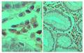
---(Pro-and-Active)-Immunohistochemistry-Paraffin-NB100-56708-img0042.jpg) | Immunohistochemistry-Paraffin: Caspase-3 Antibody (31A1067) - (Pro and Active) [NB100-56708] - IHC analysis of a formalin fixed paraffin embedded tissue section of human spleen using 1:200 dilution of Caspase-3 antibody (clone 31A1067). The staining was developed with HRP labeled anti-mouse IgG secondary antibody and DAB reagent, and nuclei of cells were counter-stained with hematoxylin. This Caspase 3 antibody generated primarily a specific cytoplasmic staining in a subset of spleenocytes with some nuclear signal in a few cells. |
---(Pro-and-Active)-Western-Blot-NB100-56708-img0040.jpg) | Western Blot: Caspase-3 Antibody (31A1067) - (Pro and Active) [NB100-56708] - Analysis using Azide/BSA FREE version of NB100-56708. Analysis of Caspase-3 in A) RAW and B) NIH 3T3 using 32 ug/ml. |
---(Pro-and-Active)-Flow-Cytometry-NB100-56708-img0043.jpg) | Flow Cytometry: Caspase-3 Antibody (31A1067) - (Pro and Active) [NB100-56708] - An intracellular stain was performed on HeLa cells with Caspase-3 Antibody (31A1067) NB100-56708 (blue) and a matched isotype control (orange). Cells were fixed with 4% PFA and then permeablized with 0.1% saponin. Cells were incubated in an antibody dilution of 5 ug/mL for 30 minutes at room temperature, followed by mouse F(ab)2 IgG (H+L) APC-conjugated secondary antibody (F0101B, R&D Systems). |
---(Pro-and-Active)-Western-Blot-NB100-56708-img0038.jpg) | Western Blot: Caspase-3 Antibody (31A1067) - (Pro and Active) [NB100-56708] - Analysis using the Azide Free version of NB100-56708. Detection of Lanes 1, 2 and 3 demonstrate the species cross-reactivity of the antibody in human, mouse and rat heart lysate, respectively. |
---(Pro-and-Active)-Immunohistochemistry-Paraffin-NB100-56708-img0027.jpg) | Immunohistochemistry-Paraffin: Caspase-3 Antibody (31A1067) - (Pro and Active) [NB100-56708] - Caspase-3 was detected in immersion fixed paraffin-embedded sections of human bladder tissue using 1:50 dilution of mouse anti-Caspase-3 monoclonal (NB100-56708, Novus Biologicals), for 1 hour at room temperature followed by anti-mouse IgG VisUCyte HRP polymer(VC001). Tissue was stained using DAB (brown) and counterstained with hematoxylin (blue). |
---(Pro-and-Active)-Immunohistochemistry-Paraffin-NB100-56708-img0029.jpg) | Immunohistochemistry-Paraffin: Caspase-3 Antibody (31A1067) - (Pro and Active) [NB100-56708] - Analysis using the Azide Free version of NB100-56708. Staining of human breast cancer stained at 4ug/ml. Localization can be cytoplasmic and nuclear. Staining in the nucleus is considered to be an indication of active Caspase-3. In most cell types and model systems, cells with active Caspase-3 are undergoing apoptosis. |
---(Pro-and-Active)-Immunohistochemistry-Paraffin-NB100-56708-img0034.jpg) | Immunohistochemistry-Paraffin: Caspase-3 Antibody (31A1067) - (Pro and Active) [NB100-56708] - Analysis using the azide free version of NB100-56708. Staining of normal colon stained 4ug/ml. Localization can be cytoplasmic and nuclear. Staining in the nucleus is considered to be an indication of active Caspase-3. In most cell types and model systems |
---(Pro-and-Active)-Immunohistochemistry-Paraffin-NB100-56708-img0035.jpg) | Immunohistochemistry-Paraffin: Caspase-3 Antibody (31A1067) - (Pro and Active) [NB100-56708] - Analysis using the Azide Free version of NB100-56708. Staining of the human thymus after heat-induced antigen retrieval at 5 ug/ml. |
追加しました。
Background
Caspases are a family of cysteine proteases that are key mediators of programmed cell death or apoptosis. The precursor form of all caspases is composed of a prodomain, and large and small catalytic subunits. The active forms of caspases are generated by several stimuli including ligand-receptor interactions, growth factor deprivation and inhibitors of cellular functions. All known caspases require cleavage adjacent to aspartates to liberate one large and one small subunit, which associate into a2b2 tetramer to form the active enzyme. Gene for Caspase-3 also known as Yama, CPP32, and apopain codes for a 32-kDa protein. Caspase-3 cleaves the death substrate poly(ADP-ribose) polymerase (PARP) to a specific 85 kDa form observed during apoptosis and is inhibitable by the CrmA protein. Other Caspase-3 substrates include DNA-PK, actin, GAS2, and procaspase-6, etc. Caspase-3 is activated by cleavage events at Asp-28/Ser-29 (between N-terminal pro-domain) and Asp-175/Ser-176 (between large and small subunits) to generate a large subunit of 17-kDa and a small subunit of 12-kDa.追加しました。
製品情報は掲載時点のものですが、価格表内の価格については随時最新のものに更新されます。お問い合わせいただくタイミングにより製品情報・価格などは変更されている場合があります。
表示価格に、消費税等は含まれていません。一部価格が予告なく変更される場合がありますので、あらかじめご了承下さい。