
抗TLR4 (76B357.1)抗体 | Anti-TLR4 (76B357.1) Antibody
掲載日情報:2018/07/09 現在Webページ番号:47209

世界最大級の抗体製品数を取り扱うNovus Biologicals社のTLR4 (76B357.1)に対する抗体(anti-TLR4 (76B357.1) | antibody TLR4 (76B357.1))です。Novus Biologicals社の抗体は数多くの学術論文で使用実績があります。
※本製品は研究用です。研究用以外には使用できません。

追加しました。
価格
[在庫・価格 :2026年07月14日 13時55分現在]
| 詳細 | 商品名 |
|
文献数 | ||||||||||||||||||||||||||||||||||||||||||||||||||||||||||||||||||||||||||||||||||
|---|---|---|---|---|---|---|---|---|---|---|---|---|---|---|---|---|---|---|---|---|---|---|---|---|---|---|---|---|---|---|---|---|---|---|---|---|---|---|---|---|---|---|---|---|---|---|---|---|---|---|---|---|---|---|---|---|---|---|---|---|---|---|---|---|---|---|---|---|---|---|---|---|---|---|---|---|---|---|---|---|---|---|---|---|---|
|
Anti-TLR4, Mouse-Mono(76B357.1) <Anti-CD284> |
|
112 | |||||||||||||||||||||||||||||||||||||||||||||||||||||||||||||||||||||||||||||||||||
|
|||||||||||||||||||||||||||||||||||||||||||||||||||||||||||||||||||||||||||||||||||||


[在庫・価格 :2026年07月14日 13時55分現在]
Anti-TLR4, Mouse-Mono(76B357.1) <Anti-CD284>
文献数: 112
- 商品コード:NB100-56566
- メーカー:NOV
- 包装:0.1mg
- 価格:¥108,000
- 在庫:無(未発注)
- 納期:3~4週間 ※※ 表示されている納期は弊社に在庫がなく、取り寄せた場合の目安納期となります。
- 法規制等:
| 説明文 | レビューあり。旧IMGENEX社 商品コード:IMG-5031A,Keywords:76B357.1|ARMD10|CD284|CD284 antigen|hTollhomolog of Drosophila toll|tlr4 76B357.1|TLR4 facs|TLR4 flow cytometry|TLR4 human|TLR4 ihc|TLR4 western blot|TOLL|toll-like receptor 4 クローン:76B357.1 Genbank No: 7099 Protein Accession No: O00206 |
||||||
|---|---|---|---|---|---|---|---|
| 別包装品 | 別包装品あり | ||||||
| 法規制等 | |||||||
| 保存条件 | -20℃ | 法規備考 | |||||
| 抗原種 | 免疫動物 | Mouse | |||||
| 交差性 | Bovine/Human/Mammal/Mouse/Porcine/Rat | 適用 | ChIP,Dot,ELISA,FCM,IC,IF,IHC,Neutralising,SDS-PAGE,Western Blot,in vitro assay | ||||
| 標識 | Unlabeled | 性状 | Protein A/G Affinity Purified | ||||
| 吸収処理 | クラス | IgG | |||||
| クロナリティ | Monoclonal | フォーマット | |||||
| 掲載カタログ |
|
||||||
| 製品記事 | Toll - Like Receptor ( TLR ) 関連抗体 Toll-Like Receptor(TLR) Screening Kit /抗 TLR 抗体 フローサイトメトリー用抗 TLR 抗体 |
||||||
| 関連記事 | |||||||
追加しました。
Image
-Western-Blot-NB100-56566-img0018.jpg) | Western Blot: TLR4 Antibody (76B357.1) [NB100-56566] - Analysis using 2 ug/ml on (A) human intestine and 6 ug/ml on (B) mouse intestine and C) rat intestine lysate. |
-Immunohistochemistry-Paraffin-NB100-56566-img0007.jpg) | Immunohistochemistry-Paraffin: TLR4 Antibody (76B357.1) [NB100-56566] - Formalin-fixed, paraffin-embedded human skin stained with at 5 ug/ml, peroxidase-conjugate and DAB chromogen. Staining of formalin-fixed tissues is enhanced by boiling tissue sections in 10 mM sodium citrate buffer, pH 6.0 for 10-20 min followed by cooling at RT for 20 min. |
-Flow-(Intracellular)-NB100-56566-img0025.jpg) | Flow (Intracellular): TLR4 Antibody (76B357.1) [NB100-56566] - Analysis using PE conjugate of NBP2-27149. An intracellular stain was performed on Jurkat cells with TLR4 antibody (76B357.1) NBP2-27149PE (blue) and an isotype control MAB004 (orange). Cells were fixed with 4% PFA and then permeablized with 0.1% saponin. Cells were incubated in an antibody dilution of 2.5 ug/mL for 30 minutes at room temperature. Both antibodies were conjugated to phycoerythrin. |
-Flow-Cytometry-NB100-56566-img0031.jpg) | Flow Cytometry: TLR4 Antibody (76B357.1) [NB100-56566] - Flow cytometric analysis of formaldehyde fixed THP-1 cells (human monocytic leukemia cells) using 2 ug/10^6 cells TLR4 antibody (clone 76B357.1) with detection employing a donkey anti-mouse IgG (H+L) cross adsorbed secondary antibody, (DyLight 488 conjugated). Isotype control samples incubated with mouse IgG2b isotype control antibody were processed in parallal under the same assay conditions. |
-Flow-Cytometry-NB100-56566-img0024.jpg) | Flow Cytometry: TLR4 Antibody (76B357.1) [NB100-56566] - Analysis using the Alexa Fluor (R) 647 conjugate of NBP2-27149. TLR4 expression on monocytes from human peripheral blood: PBMC were stained, in a 2 color flow test, with CD14 PE this antibody and 1 ug of either isotype control (Left, ) or TLR4-Alexa Fluor 647 (right). PPI negative, CD14+ cells were gated for analysis. |
-Flow-Cytometry-NB100-56566-img0009.jpg) | Flow Cytometry: TLR4 Antibody (76B357.1) [NB100-56566] - Flow analysis of 10^6 ThP1 cells using 5 ug. Shaded histogram shows ThP1 cells without anti-TLR4 antibody; green represents isotype control; red represents anti-TLR4 antibody. |
-Flow-(Cell-Surface)-NB100-56566-img0028.jpg) | Flow (Cell Surface): TLR4 Antibody (76B357.1) [NB100-56566] - Analysis using the FITC conjugate of NBP2-27149. Staining of cell surface TLR4 in human monocyte-derived dendritic cells using this antibody at 1:200 (2.5 ug). Black and green open histograms represent unstained cells and negative control (anti-human CD14 |
-Immunohistochemistry-Paraffin-NB100-56566-img0013.jpg) | Immunohistochemistry-Paraffin: TLR4 Antibody (76B357.1) [NB100-56566] - Analysis of formalin-fixed paraffin-embedded tissue section of normal human skin at 5 ug/ml. Membrane-cytoplasmic immunopositivity of TLR4 was primarily observed in the pigmented basel cells and the adjacent keratinocytes in the epidermal layer. |
-Immunohistochemistry-Frozen-NB100-56566-img0032.jpg) | Immunohistochemistry-Frozen: TLR4 Antibody (76B357.1) [NB100-56566] - This image is TLR4(green) and nucleus(blue) at area postrema of the adult male mouse brai n, x20 magnification. Primary antibody diluted 1:500. This image was submitted via customer Review. |
-Flow (Intracellular)-NB100-56566-img0033.jpg) | Flow (Intracellular): TLR4 Antibody (76B357.1) [NB100-56566] - An intracellular stain was performed on Jurkat cells with TLR4 antibody (76B357.1) NBP2-27149AF647 (blue) and a matched isotype control (orange). Cells were fixed with 4% PFA and then permeablized with 0.1% saponin. Cells were incubated in an antibody dilution of 2.5 ug/mL for 30 minutes at room temperature. Both antibodies were conjugated to Alexa Fluor 647. Image from the Alexa Fluor 647 version of this antibody. |
-Immunohistochemistry-Paraffin-NB100-56566-img0010.jpg) | Immunohistochemistry-Paraffin: TLR4 Antibody (76B357.1) [NB100-56566] - Analysis of formalin-fixed paraffin-embedded tissue section of normal human breast at 5 ug/ml. The ductal/acinar epithelial cells and the myoepithelial cells showed specific membrane-cytoplasmic positivity for TLR4. Mild immunoreactivity of TLR4 was observed in blood vessels and the intra-lobular connective tissue also. |
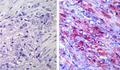
-Immunohistochemistry-Paraffin-NB100-56566-img0011.jpg) | Immunohistochemistry-Paraffin: TLR4 Antibody (76B357.1) [NB100-56566] - Analysis of formalin-fixed paraffin-embedded tissue section of human esophageal squamous cell carcinoma (SCC) using 5 ug/ml. Membrane-cytoplasmic immunopositivity was observed in SCC cells while the tumor stroma was largely negative for TLR4. |
-Immunohistochemistry-Paraffin-NB100-56566-img0014.jpg) | Immunohistochemistry-Paraffin: TLR4 Antibody (76B357.1) [NB100-56566] - Analysis of formalin-fixed paraffin-embedded tissue section of malignant stromal tumor of small intestine from human at 5 ug/ml. The representative image shows a membrane-cytoplasmic staining pattern of TLR4 in the cancer cells. |
-Immunohistochemistry-Paraffin-NB100-56566-img0015.jpg) | Immunohistochemistry-Paraffin: TLR4 Antibody (76B357.1) [NB100-56566] - Analysis of Rat's salivary gland tissue section at 1:100 dilution. The antibody generated a membrane-cytoplasmic staining in the tissue with stronger signal in ductal epithelial cells. |
-Immunohistochemistry-Paraffin-NB100-56566-img0016.jpg) | Immunohistochemistry-Paraffin: TLR4 Antibody (76B357.1) [NB100-56566] - Analysis of TLR4 in paraffin-embedded formalin-fixed human colon tissue using an isotype control (top) and NB100-56566 (bottom) at 5 ug/ml. |
-Immunohistochemistry-Paraffin-NB100-56566-img0017.jpg) | Immunohistochemistry-Paraffin: TLR4 Antibody (76B357.1) [NB100-56566] - Formalin-fixed, paraffin-embedded human testis tissue stained using 5 ug/ml. |
-Flow-Cytometry-NB100-56566-img0006.jpg) | Flow Cytometry: TLR4 Antibody (76B357.1) [NB100-56566] - Surface staining of mouse splenocytes using TLR4 antibody (0.5 ug/10^6 cells) and a PE-conjugated secondary this antibody. Shaded histogram represents cells without TLR4 antibody; green represents isotype control (BD Pharmingen) red represents TLR4 antibody. |
-Flow-Cytometry-NB100-56566-img0019.jpg) | Flow Cytometry: TLR4 Antibody (76B357.1) [NB100-56566] - Analysis of FITC conjugate of NB100-56566. Cell surface TLR4 in human monocyte-derived dendritic cells using this antibody at 1:200 (2.5 ug). Black and green open histograms represent unstained cells and negative control (anti-human CD14 FITC, respectivel |
-Flow-Cytometry-NB100-56566-img0026.jpg) | Flow Cytometry: TLR4 Antibody (76B357.1) [NB100-56566] - Analysis using the FITC conjugate of NBP2-27149. TLR4 expression on monocytes from human peripheral blood: PBMC were stained, in a 2 color flow test, with CD14 PE and 1 ug of either isotype control (Left, ) or TLR4-FITC (Right, this antibody. PPI negative, CD14+ cells were gated for analysis. |
追加しました。
Background
The Toll-like receptor (TLR) family in mammal comprises a family of transmembrane proteins characterized by multiple copies of leucine rich repeats in the extracellular domain and IL-1 receptor motif in the cytoplasmic domain. Like its counterparts in Drosophila, TLRs signal through adaptor molecules. The TLR family is a phylogenetically conserved mediator of innate immunity that is essential for microbial recognition. Ten human homologs of TLRs (TLR1-10) have been described. Among this family of receptors, TLR2 and TLR4 have been most studied. These studies have suggested that TLR2 and TLR4 may serve as potential main mediators of LPS signaling. The TLR4 cDNA codes for a protein consisting of 799 amino acids with approximate molecular weight of 88 kDa.追加しました。
製品情報は掲載時点のものですが、価格表内の価格については随時最新のものに更新されます。お問い合わせいただくタイミングにより製品情報・価格などは変更されている場合があります。
表示価格に、消費税等は含まれていません。一部価格が予告なく変更される場合がありますので、あらかじめご了承下さい。