
抗HIF-1 alpha抗体 | Anti-HIF-1 alpha Antibody
掲載日情報:2018/07/09 現在Webページ番号:46964

世界最大級の抗体製品数を取り扱うNovus Biologicals社のHIF-1 alphaに対する抗体(anti-HIF-1 alpha | antibody HIF-1 alpha)です。Novus Biologicals社の抗体は数多くの学術論文で使用実績があります。
※本製品は研究用です。研究用以外には使用できません。

追加しました。
価格
[在庫・価格 :2026年07月17日 00時01分現在]
| 詳細 | 商品名 |
|
文献数 | ||||||||||||||||||||||||||||||||||||||||||||||||||||||||||||||||||||||||||||||||||
|---|---|---|---|---|---|---|---|---|---|---|---|---|---|---|---|---|---|---|---|---|---|---|---|---|---|---|---|---|---|---|---|---|---|---|---|---|---|---|---|---|---|---|---|---|---|---|---|---|---|---|---|---|---|---|---|---|---|---|---|---|---|---|---|---|---|---|---|---|---|---|---|---|---|---|---|---|---|---|---|---|---|---|---|---|---|
|
Anti-HIF-1α, Rabbit-Poly <Anti-Hypoxia-Inducible Factor 1α> |
|
244 | |||||||||||||||||||||||||||||||||||||||||||||||||||||||||||||||||||||||||||||||||||
|
|||||||||||||||||||||||||||||||||||||||||||||||||||||||||||||||||||||||||||||||||||||


[在庫・価格 :2026年07月17日 00時01分現在]
Anti-HIF-1α, Rabbit-Poly <Anti-Hypoxia-Inducible Factor 1α>
文献数: 244
- 商品コード:NB100-449
- メーカー:NOV
- 包装:0.1ml
- 価格:¥108,000
- 在庫:無(未発注)
- 納期:3~4週間 ※※ 表示されている納期は弊社に在庫がなく、取り寄せた場合の目安納期となります。
- 法規制等:
| 説明文 | レビューあり。Simple Western対応抗体。抗原:ヒトhypoxia-inducible factor 1, alpha subunit C-terminal(775-826aa)付近の配列を持つ合成ペプチド,Keywords:ARNT-interacting protein|Basic-helix-loop-helix-PAS protein MOP1|BHLHE78|Class E basic helix-loop-helix protein 78|HIF-1 alpha|HIF1A|HIF-1-alpha|HIF1-alpha|hypoxia inducible factor 1|alpha subunit (basic helix-loop-helix transcriptionfactor) Genbank No: 3091 Protein Accession No: Q16665 |
||||||
|---|---|---|---|---|---|---|---|
| 別包装品 | 別包装品あり | ||||||
| 法規制等 | |||||||
| 保存条件 | 4℃,凍結禁止 | 法規備考 | |||||
| 抗原種 | Human | 免疫動物 | Rabbit | ||||
| 交差性 | Canine/Chicken/Goat/Human/Monkey/Mouse/Primate/Rat | 適用 | ChIP,ELISA,FCM,IC,IF,IHC,IP,Simple Western,Western Blot | ||||
| 標識 | Unlabeled | 性状 | Antigen Affinity Purified | ||||
| 吸収処理 | クラス | IgG | |||||
| クロナリティ | Polyclonal | フォーマット | |||||
| 掲載カタログ |
|
||||||
| 製品記事 | 抗HIF抗体(Anti-HIF-1/HIF-2 antibody) |
||||||
| 関連記事 | |||||||
追加しました。
Image
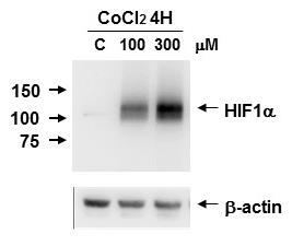 | Biological Strategies Validation. Western Blot: HIF-1 alpha Antibody [NB100-449] - HIF-1 alpha induction on Caki-1 cell lysate using CoCl2. Image from verified customer review. |
 | Simple Western: HIF-1 alpha Antibody [NB100-449] - Simple Western lane view shows a specific band for HIF-1 alpha in 0.5 mg/ml of Hypoxic HeLa lysate. This experiment was performed under reducing conditions using the 12-230 kDa separation system. |
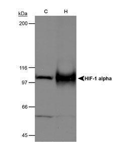 | Western Blot: HIF-1 alpha Antibody [NB100-449] - Detection of mouse HIF1-alpha on hypoxia treated MEFs |
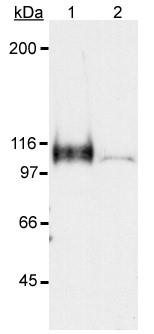 | Western Blot: HIF-1 alpha Antibody [NB100-449] - Detection of HIF-1 alpha in a hypoxic sample. Lane 1: CoCl2 treated Cos-7 nuclear extract (hypoxic). Lane 2: Untreated Cos-7 nuclear extract (normoxic). |
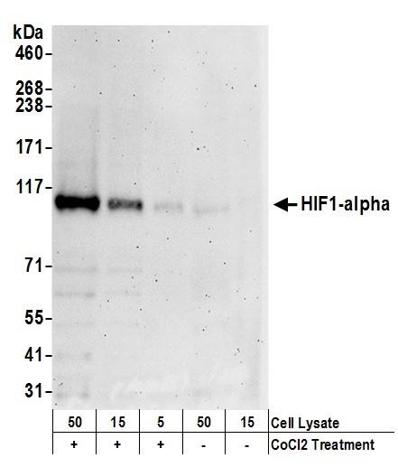
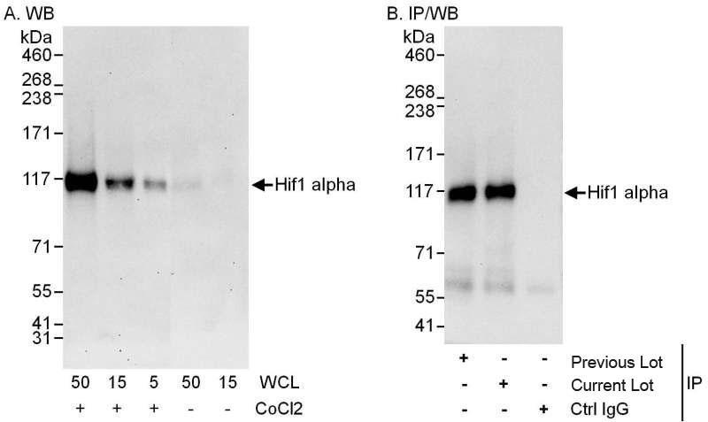 | Independent Antibodies Validation. Western Blot: HIF-1 alpha Antibody [NB100-449] - Detection of Human HIF1 alpha by Western Blot and Immunoprecipitation. Samples: Whole cell lysate (5, 15 and 50 ug for WB; 1 mg for IP, 20% of IP loaded) from HeLa cells that were either treated with cobalt chloride (+; 200 mcM) or mock treated (-). Antibodies: Affinity purified rabbit anti-HIF1 alpha antibody used for WB at 0.1 ug/ml (A) and 1 ug/ml (B) and used for IP at 3 ug/mg lysate. HIF1 alpha was also immunoprecipitated by a previous lot of this antibody. Detection: Chemiluminescence with exposure times of 30 seconds (A) and 10 seconds (B). |
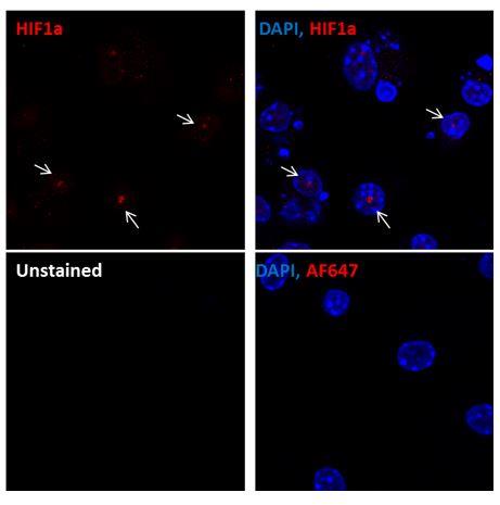
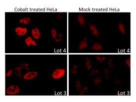 | Biological Strategies Validation. Immunocytochemistry/Immunofluorescence: HIF-1 alpha Antibody [NB100-449] - Formaldehyde-fixed asynchronous HeLa cells. |
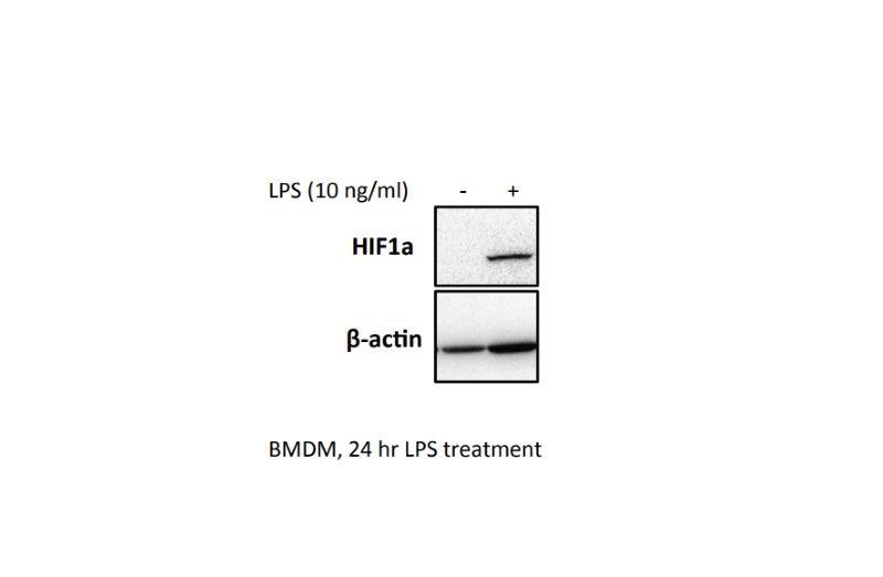 | Western Blot: HIF-1 alpha Antibody [NB100-449] - BMDM were seeded at 0.5x10^6 overnight. Cells were treated with 10 ng/ml LPS for 24 hrs, and a western blot was performed. This image was submitted via customer Review. |
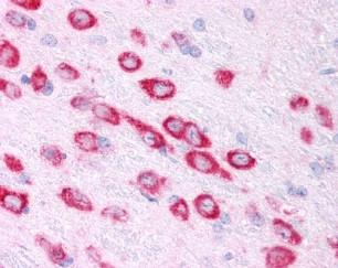 | Immunohistochemistry: HIF-1 alpha Antibody [NB100-449] - Mouse Brain, Neurons 40X |
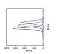
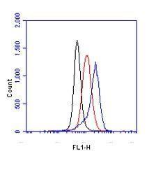 | Flow Cytometry: HIF-1 alpha Antibody [NB100-449] - Hela cells were treated for 15 hrs with 200uM CoCl2, fixed in PFA, and permeabilized in 90% MeOH. 1 X 10^6 cells were stained with 0.125ug anti- HIF-alpha and secondary FITC-conjugated goat anti-rabbit (in a 150ul reaction). Black- treated, anti-KLH control IgG; Red- untreated, anti-HIF1-alpha; Blue- treated, anti-HIF1-alpha. |
 | Western Blot: HIF-1 alpha Antibody [NB100-449] - Homogenate from pig (lanes 1 and 2) or rabbit (lane 4) aorta or lysate from cultured rat aortic smooth muscle cells (lane 3). Antibody: Affinity purified rabbit anti-SERCA2 used at 1 ug/ml (lanes 2 and 4) or 0.4 ug/ml (lane 3) for WB and 2 ug/mg lysate for IP or control (ctrl) monoclonal anti-SERCA2 (lanes 1 and 4) used at 1 ug/ml for WB. Detection: Chemiluminescence. |
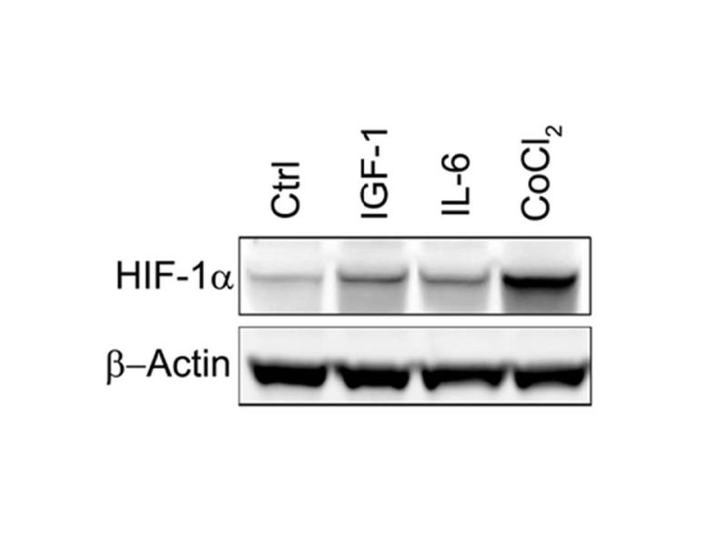 | Biological Strategies Validation. Western Blot: HIF-1 alpha Antibody [NB100-449] - Analysis of HIF-1 alpha in human myeloma cell lysate using anti-HIF-1 alpha. Cells were untreated or treated with IGF-1, IL-6 or CoCl2. Image from verified customer review. |
 | Biological Strategies Validation. Western Blot: HIF-1 alpha Antibody [NB100-449] - Detection of Human HIF1 alpha by Western Blot. Samples: Whole cell lysate (5, 15 and 50 ug) from HeLa cells that were treated with cobalt chloride (+; 200 mcM) or mock treated (-). Antibodies: Affinity purified rabbit anti-HIF1 alpha antibody NB100-449 used for WB at 0.1 ug/ml. Detection: Chemiluminescence with exposure times of 30 seconds. |
 | Immunoprecipitation: HIF-1 alpha Antibody [NB100-449] - Analysis in HEK293 cells. Image courtesy of anonymous customer review. |
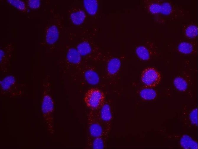 | Proximity Ligation Assay: HIF-1 alpha Antibody [NB100-449] - Secondary-conjugate Duolink II PLA in Hela cells. goat anti-human MCM2 (NB100-244) and rabbit anti-human HIF1-alpha (NB100-449). Image merged from DAPI (2ms) and Texas Red (200ms) exposures, 40X magnification. |
 | Proximity Ligation Assay: HIF-1 alpha Antibody [NB100-449] - Secondary-conjugate Duolink II PLA in Hela cells. goat anti-human MCM7 (NB100-252) and rabbit anti-human HIF1-alpha (NB100-449). Image merged from DAPI (2ms) and Texas Red (200ms) exposures, 40X magnification. |
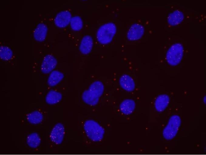 | Proximity Ligation Assay: HIF-1 alpha Antibody [NB100-449] - Secondary-conjugate Duolink II PLA in Hela cells. goat anti-human MCM3 (NB100-249) and rabbit anti-human HIF1-alpha (NB100-449). Image merged from DAPI (2ms) and Texas Red (200ms) exposures, 40X magnification. |
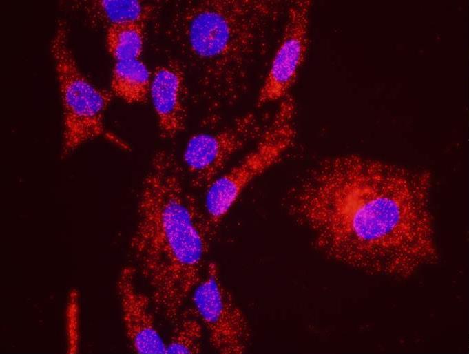
 | Immunofluorescence: HIF-1 alpha Antibody [NB100-449] - Murine primary bone marrow derived macrophages stained with HIF1-alpha antibody (red). Nuclei were counterstained with Dapi (blue). Image from verified customer review. |
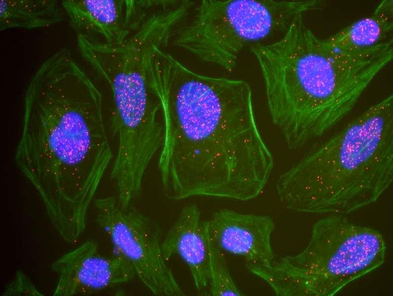 | Proximity Ligation Assay: HIF-1 alpha Antibody [NB100-449] - Shows representative results for PLA using three color fluorescence, including DAPI stained nuclei (blue), phalloidin stained cytoplasmic F-actin (green), and PLA positive signal (red). |
追加しました。
Background
Hypoxia contributes significantly to the pathophysiology of major categories of human disease, including myocardial and cerebral ischemia, cancer, pulmonary hypertension, congenital heart disease and chronic obstructive pulmonary disease. HIF-1 is a nuclear protein involved in mammalian oxygen homeostasis. This occurs as a posttranslational modification by prolyl hydroxylation. HIF-1 is a heterodimer composed of HIF-1 alpha and HIF-1 beta subunits. Both subunits are constantly translated. However, under normoxic conditions, human HIF-1 alpha is hydroxylated at Pro402 or Pro564 by a set of HIF prolyl hydroxylases, is polyubiquinated, and eventually degraded in proteosomes. Under hypoxic conditions, the lack of hydroxylation prevents HIF degradation and increases transcriptional activity. Therefore, the concentration of HIF-1 alpha increases in the cell. In contrast, HIF-1 beta remains stable under either condition. HIF hydroxylases provide insight into hypoxic cell responses, which may be used to help isolate therapeutic targets.追加しました。
製品情報は掲載時点のものですが、価格表内の価格については随時最新のものに更新されます。お問い合わせいただくタイミングにより製品情報・価格などは変更されている場合があります。
表示価格に、消費税等は含まれていません。一部価格が予告なく変更される場合がありますので、あらかじめご了承下さい。