
HOME > 試薬 > 抗体 > 一次抗体 > 抗LC3B抗体 | Anti-LC3B Antibody
抗LC3B抗体 | Anti-LC3B Antibody
掲載日情報:2018/07/09 現在Webページ番号:46612

世界最大級の抗体製品数を取り扱うNovus Biologicals社のLC3Bに対する抗体(anti-LC3B | antibody LC3B)です。Novus Biologicals社の抗体は数多くの学術論文で使用実績があります。
※本製品は研究用です。研究用以外には使用できません。

追加しました。
価格
[在庫・価格 :2026年07月15日 20時55分現在]
| 詳細 | 商品名 |
|
文献数 | ||||||||||||||||||||||||||||||||||||||||||||||||||||||||||||||||||||||||||||||||||
|---|---|---|---|---|---|---|---|---|---|---|---|---|---|---|---|---|---|---|---|---|---|---|---|---|---|---|---|---|---|---|---|---|---|---|---|---|---|---|---|---|---|---|---|---|---|---|---|---|---|---|---|---|---|---|---|---|---|---|---|---|---|---|---|---|---|---|---|---|---|---|---|---|---|---|---|---|---|---|---|---|---|---|---|---|---|
|
Anti-LC3, Human, Rabbit-Poly |
|
1231 | |||||||||||||||||||||||||||||||||||||||||||||||||||||||||||||||||||||||||||||||||||
|
|||||||||||||||||||||||||||||||||||||||||||||||||||||||||||||||||||||||||||||||||||||
|
Anti-LC3, Rabbit-Poly |
|
285 | |||||||||||||||||||||||||||||||||||||||||||||||||||||||||||||||||||||||||||||||||||
|
|||||||||||||||||||||||||||||||||||||||||||||||||||||||||||||||||||||||||||||||||||||


[在庫・価格 :2026年07月15日 20時55分現在]
Anti-LC3, Human, Rabbit-Poly
文献数: 1231
- 商品コード:NB100-2220
- メーカー:NOV
- 包装:0.1ml
- 価格:¥110,000
- 在庫:1個
- 納期:3~4週間 ※※ 表示されている納期は弊社に在庫がなく、取り寄せた場合の目安納期となります。
- 法規制等:
| 説明文 | レビューあり。Simple Western対応抗体。抗原:ヒトLC3(aa 1~100),Keywords:anti-LC3B|atg8f Autophagy-related protein LC3 B|Autophagy-related ubiquitin-like modifier LC3 B|LC3B|lc3b autophagy marker|LC3B ihc|LC3B immunohistochemistry|LC3B immunoprecipitation|LC3B western blot|lc3-i|ii|MAP1 light chain 3-like protein 2 Genbank No: 81631 Protein Accession No: Q9GZQ8 |
||||||
|---|---|---|---|---|---|---|---|
| 別包装品 | 別包装品あり | ||||||
| 法規制等 | |||||||
| 保存条件 | -20℃ | 法規備考 | |||||
| 抗原種 | Human | 免疫動物 | Rabbit | ||||
| 交差性 | Alligator/Avian/Bacteria/Bovine/Canine/Chicken/Chinese Hamster/Golden Syrian Hamster/Guinea Pig/Hamster/Human/Invertebrate/Monkey/Mouse/Porcine/Primate/Rabbit/Rat/Zebrafish | 適用 | ChIP,ELISA,FCM,IC,IF,IHC,IP,Immunoblotting,Proximity Ligation Assay,SDS-PAGE,Simple Western,Western Blot | ||||
| 標識 | Unlabeled | 性状 | Antigen Affinity Purified | ||||
| 吸収処理 | クラス | IgG | |||||
| クロナリティ | Polyclonal | フォーマット | |||||
| 掲載カタログ |
|
||||||
| 製品記事 | 抗LC3B抗体 | Anti- LC3B antibody 抗LC3抗体/抗LC3関連転写因子抗体 抗LC3抗体 オートファジーとLC3研究用試薬 オルガネラ(細胞小器官)マーカー抗体 |
||||||
| 関連記事 | |||||||
Anti-LC3, Rabbit-Poly
文献数: 285
- 商品コード:NB100-2220SS
- メーカー:NOV
- 包装:0.025ml
- 価格:¥55,000
- 在庫:無(未発注)
- 納期:3~4週間 ※※ 表示されている納期は弊社に在庫がなく、取り寄せた場合の目安納期となります。
- 法規制等:
| 説明文 | Simple Western対応抗体。抗原:ヒトLC3(aa 1~100),Keywords:ATG8F|Autophagy-related protein LC3 B|Autophagy-related ubiquitin-like modifier LC3 B|LC3B|lc3-i|ii|MAP1 light chain 3-like protein 2|MAP1A/1BLC3|MAP1A/MAP1B LC3 B|MAP1ALC3|microtubule-associated protein 1 light chain 3 betaMAP1A/MAP1B light chain 3 B Genbank No: 81631 Protein Accession No: Q9GZQ8 |
||||||
|---|---|---|---|---|---|---|---|
| 別包装品 | 別包装品あり | ||||||
| 法規制等 | |||||||
| 保存条件 | -20℃ | 法規備考 | |||||
| 抗原種 | Human | 免疫動物 | Rabbit | ||||
| 交差性 | Alligator/Avian/Bacteria/Bovine/Canine/Chicken/Chinese Hamster/Golden Syrian Hamster/Guinea Pig/Hamster/Human/Invertebrate/Monkey/Mouse/Porcine/Primate/Rabbit/Rat/Zebrafish | 適用 | ChIP,ELISA,FCM,IC,IF,IHC,IP,Immunoblotting,Proximity Ligation Assay,SDS-PAGE,Simple Western,Western Blot | ||||
| 標識 | Unlabeled | 性状 | Antigen Affinity Purified | ||||
| 吸収処理 | クラス | IgG | |||||
| クロナリティ | Polyclonal | フォーマット | |||||
| 掲載カタログ |
ニュース2018年2月15日号 p.32
|
||||||
| 製品記事 | 抗LC3B抗体 | Anti- LC3B antibody 抗LC3抗体/抗LC3関連転写因子抗体 抗LC3抗体 オートファジーとLC3研究用試薬 |
||||||
| 関連記事 | |||||||
追加しました。
Image
 | Genetic Strategies Validation. Knockout Validated: LC3B Antibody [NB100-2220] - Western blot shows lysates of HeLa human cervical epithelial carcinoma parental cell line and LC3B knockout HeLa cell line (KO) untreated (-) or treated (+) with 50 uM Chloroquine for 18 hours. PVDF membrane was probed with 0.5 ug/mL of Rabbit Anti-LC3B Monoclonal Antibody (Catalog # NB100-2220) followed by HRP-conjugated Anti-Rabbit IgG Secondary Antibody (Catalog# HAF008). A specific band was detected for LC3B at approximately 15 kDa (as indicated) in the parental HeLa cell line, but is not detectable in the knockout HeLa cell line. GAPDH is shown as a loading control. This experiment was conducted under reducing conditions. |
 | Genetic Strategies Validation. Knockout Validated: LC3B Antibody [NB100-2220] - LC3B/MAP1 [NB100-2220] - Detection of LC3 in mouse ES cell lysates. Atg5-/- ES cells from Dr. Noboru Mizushima [Mizushima, N. et al. J. Cell Biol. 152 (2001)] Photo courtesy of Dr. Beth Levine, UT SW Medical Center. |
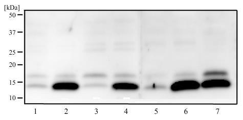 | Biological Strategies Validation. Western Blot: LC3B Antibody [NB100-2220] - LC3B/MAP1 [NB100-2220] - Western blot analysis of HeLa (1), HeLa + CQ (2), SHSY5Y (3), SHSY5Y +CQ (4), A431 (5), A431 +CQ (6) and Ntera2 (7) using LC3 antibody at 2 ug/ml. |
 | Biological Strategies Validation. Western Blot: LC3B Antibody [NB100-2220] - shows lysates of mouse NIH3T3 and rat PC-12 cell lines untreated (-) or treated (+) with Chloroquine. PVDF membrane was probed with 0.5 ug/mL rabbit anti-LC3B polyclonal Antibody (NB100-2220, Novus Biologicals), followed by 1:2000 dilution of goat anti-rabbit IgG secondary antibody. |
 | Western Blot: LC3B Antibody [NB100-2220] - Analysis of LC3 in Huh-7 and SMMC-7721 cells using anti-LC3B antibody. Image from verified customer review. |
 | Western Blot: LC3B Antibody [NB100-2220] - Analysis using the Biotin conjugate of NB100-2220. Image from verified customer review. |
 | Immunohistochemistry-Paraffin: LC3B Antibody [NB100-2220] - Human ovarian Cancer tissue stained using heat mediated antigen retrieval in pH 6.0 citrate buffer at 1:200 dilution. Image provided by verified customer review. |
 | Immunocytochemistry/Immunofluorescence: LC3B Antibody [NB100-2220] - LC3B detected in immersion fixed HeLa human cervical epithelial carcinoma cell line treated with Chloroquine using 1ug/mL rabbit anti-LC3B polyclonal (NB100-2220, Novus Biologicals). Cells were stained using donkey anti-rabbit IgG-NL557 and counterstained with DAPI (blue). |
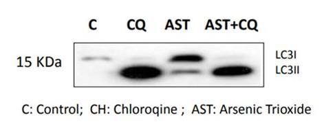 | Genetic Strategies Validation. Western Blot: LC3B Antibody [NB100-2220] - WB detection of LC3I and LC3II in mouse cochlea cell line SV-K1. Cells were treated with chloroquine (1uM), and As2O3 (1uM) for 24 hrs. This image was submitted via customer review. |
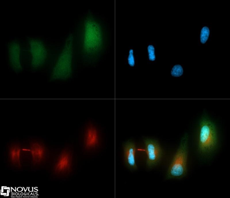 | Immunocytochemistry/Immunofluorescence: LC3B Antibody [NB100-2220] - LC3 antibody was tested in HeLa cells with Dylight 488 (green). Nuclei and alpha-tubulin were counterstained with DAPI (blue) and Dylight 550 (red). |
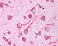
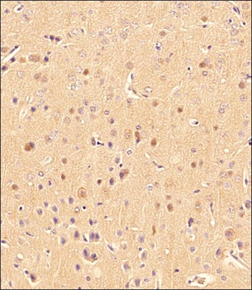
 | Immunohistochemistry: LC3B Antibody [NB100-2220] - Analysis using the Biotin conjugate of NB100-2220. Staining of brain, cerebral cortex, neurons with cell processes. |
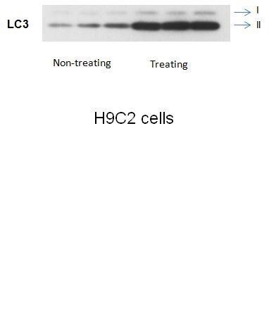 | Western Blot: LC3B Antibody [NB100-2220] - Analysis of LC3 in H9C2 cells. |
 | Western Blot: LC3B Antibody [NB100-2220] - Analysis using the DyLight 550 conjugate of NB100-2220. Detection of LC3 in pancreatic cancer cells. |
 | Western Blot: LC3B Antibody [NB100-2220] - Detection of LC3 in mouse HL-1 cell lysate. Photo courtesy of product review by verified customer. |
 | Western Blot: LC3B Antibody [NB100-2220] - Macrophage lysates probed with anti-LC3 antibody. This image was submitted via customer Review. |
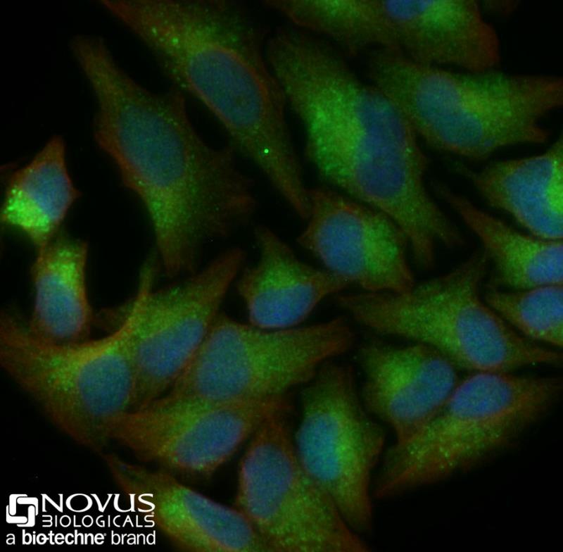 | Immunocytochemistry/Immunofluorescence: LC3B Antibody [NB100-2220] - HeLa cells were treated with 50uM CQ overnight, fixed for 10 minutes using 10% formalin and then permeabilized for 5 minutes using 1X PBS + 0.05% Triton-X100. The cells were incubated with anti-LC3B at 2 ug/ml overnight at 4C and detected with an anti-rabbit Dylight 488 (Green) at a 1:500 dilution. Alpha tubulin (DM1A) NB100-690 was used as a co-stain at a 1:1000 dilution and detected with an anti-mouse Dylight 550 (Red) at a 1:500 dilution. Nuclei were counterstained with DAPI (Blue). Cells were imaged using a 40X objective. |
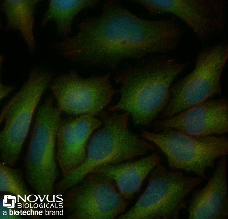 | Immunocytochemistry/Immunofluorescence: LC3B Antibody [NB100-2220] - Untreated HeLa cells were fixed for 10 minutes using 10% formalin and then permeabilized for 5 minutes using 1X PBS + 0.05% Triton-X100. The cells were incubated with anti-LC3B at 2 ug/ml overnight at 4C and detected with an anti-rabbit Dylight 488 (Green) at a 1:500 dilution. Alpha tubulin (DM1A) NB100-690 was used as a co-stain at a 1:1000 dilution and detected with an anti-mouse Dylight 550 (Red) at a 1:500 dilution. Nuclei were counterstained with DAPI (Blue). Cells were imaged using a 40X objective. |
 | Immunohistochemistry: LC3B Antibody [NB100-2220] - IHC of rat brain from confirmed customer review. |
 | Immunohistochemistry-Paraffin: LC3B Antibody [NB100-2220] - IHC analysis of a formalin fixed and paraffin embedded tissue section of mouse brain using 1:200 dilution of rabbit anti-LC3 antibody. The specific signal of LC3 was detected using HRP-conjugated secondary antibody with DAB reagent, and nuclei of cells were counterstained using hematoxylin. This LC3 antibody generated a low to moderate levels of cytoplasmic staining in the glial cells. The neurons depicted a moderate to strong staining for LC3 in their cytoplasm. |
 | Immunohistochemistry-Frozen: LC3B Antibody [NB100-2220] - LC3 accumulation in muscle fibers from patients with PAD (row B - E). Normal fibers from a Non-PAD sample (row A). Scale bar = 100 uM. This image was submitted via customer Review. |
 | Simple Western: LC3B Antibody [NB100-2220] - Simple Western lane view shows a specific band for LC3 in 0.5 mg/ml of Neuro2A lysate. This experiment was performed under reducing conditions using the 12-230 kDa separation system. |
追加しました。
Background
LC3 (microtubule-associated protein light chain 3), the most studied autophagy biomarker, was originally identified as a subunit of microtubule-associated proteins 1A and 1B (MAP1LC3) and was later found to contain similarity to yeast protein Apg8/Aut7/Cvt5. Distributed ubiquitously in eukaryotes, LC3 is expressed as 3 splice variants/isoforms (LC3A, LC3B and LC3C) which undergo post-translational processing, wherein, the unprocessed form of LC3 is proteolytically cleaved by Atg4 protease to form LC3-I with carboxyterminal exposed glycine. During autophagy, this exposed glycine of LC3-I is conjugated by Atg7 (an E1-like activity), Atg3 (an E2-like conjugating activity) and by Atg12-Atg5-Atg16L multimers (E3-like ligase activity) to phosphatidylethanolamine (PE) moiety for generating LC3-II. The lipophilic character of PE group facilitates LC3-II insertion into autophagosomes membranes, and as a result LC3-II is degraded when autophagosomes fuse with lysosomes to form autolysosomes for lysus of intra-autophagosomal components by lysosomal hydrolases. Conversion of LC3I to LC3II when correlated with autophagosome numbers is considered as the best marker of autophagy because LC3-II is the only well-characterized protein which specifically localize to autophagic structures throughout autophagy (from phagophore to lysosomal degradation). LC3 is a great tool in research as autophagy is implicated in numerous physiological/pathological processes including responses to exercise/aging, cancer, metabolic and neurodegenerative disorders, and cardiovascular/pulmonary diseases.追加しました。
製品情報は掲載時点のものですが、価格表内の価格については随時最新のものに更新されます。お問い合わせいただくタイミングにより製品情報・価格などは変更されている場合があります。
表示価格に、消費税等は含まれていません。一部価格が予告なく変更される場合がありますので、あらかじめご了承下さい。