
抗β-Tubulin抗体(Anti-β-Tubulin, Neuronal Class Ⅲ, Mouse-Mono antibody)
掲載日情報:2018/11/26 現在Webページ番号:30000

β-Tubulinに対する抗体(Anti-β-Tubulin, Neuronal Class Ⅲ, Mouse-Mono )です。
※ 本製品は研究用です。研究用以外には使用できません。
カートに商品を
追加しました。
追加しました。
価格
[在庫・価格 :2026年06月02日 00時00分現在]
※ 表示されている納期は弊社に在庫が無く、取り寄せた場合の納期目安となります。
| 詳細 | 商品名 |
|
文献数 | ||
|---|---|---|---|---|---|
|
Anti-β-Tubulin, Neuronal Class III, Mouse-Mono(Tuj-1) |
|
170 | |||
|
Anti-Neuron-specific beta-III Tubulin MAb (Clone TuJ-1) |
|
161 | |||


[在庫・価格 :2026年06月02日 00時00分現在]
※ 表示されている納期は弊社に在庫が無く、取り寄せた場合の納期目安となります。
Anti-β-Tubulin, Neuronal Class III, Mouse-Mono(Tuj-1)
文献数: 170
- 商品コード:MAB1195
- メーカー:RSD
- 包装:100μg
- 価格:¥78,000
- 在庫:1個
- 納期:10日程度 ※※ 表示されている納期は弊社に在庫がなく、取り寄せた場合の目安納期となります。
- 法規制等:
Anti-Neuron-specific beta-III Tubulin MAb (Clone TuJ-1)
文献数: 161
- 商品コード:MAB1195-SP
- メーカー:RSD
- 包装:25μg
- 価格:¥28,000
- 在庫:無(未発注)
- 納期:2~3週間 ※※ 表示されている納期は弊社に在庫がなく、取り寄せた場合の目安納期となります。
- 法規制等:
| 説明文 | Simple Western対応抗体。※受注発注品。形状:溶液または凍結乾燥 別名:Beta3-tubulin クローン:TuJ-1 Genbank No: 10381 |
||||||
|---|---|---|---|---|---|---|---|
| 別包装品 | 別包装品あり | ||||||
| 法規制等 | |||||||
| 保存条件 | -20℃ | 法規備考 | |||||
| 抗原種 | 免疫動物 | Mouse | |||||
| 交差性 | Multi-species | 適用 | IC,IHC,Simple Western,Western Blot | ||||
| 標識 | Unlabeled | 性状 | Protein A/G Affinity Purified | ||||
| 吸収処理 | クラス | IgG | |||||
| クロナリティ | Monoclonal | フォーマット | |||||
| 掲載カタログ |
|
||||||
| 製品記事 | 使いっきり抗体 |
||||||
| 関連記事 | |||||||
カートに商品を
追加しました。
追加しました。
Product Details
| Label | Unconjugated |
|---|---|
| Immunogen | Rat brain-derived microtubules |
| Source | Monoclonal Mouse IgG2A Clone # TuJ-1 |
| Purification | Protein A or G purified from hybridoma culture supernatant |
| Specificity | Detects mammalian and chicken neuron-specific beta -III tubulin but not other beta -tubulin isotypes in Western blots. |
カートに商品を
追加しました。
追加しました。
Applications and Data
| Recommended Concentration | Sample | |
| Western Blot | 0.2 µg/mL | See below |
| Simple Western | 10 µg/mL | See below |
| Immunocytochemistry | 8-25 µg/mL | See below |
| Western Blot | |
|---|---|
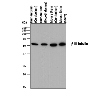 | Detection of Human and Mouse beta ‑III Tubulin by Western Blot. Western blot shows lysates of human brain (cerebellum) tissue, human brain (hypothalamus) tissue, mouse brain (cerebellum) tissue, and mouse brain (stem) tissue. PVDF membrane was probed with 0.2 µg/mL of Mouse Anti-Neuron-specific beta ‑III Tubulin Monoclonal Antibody (Catalog # MAB1195) followed by HRP-conjugated Anti-Mouse IgG Secondary Antibody (Catalog # HAF018). A specific band was detected for beta ‑III Tubulin at approximately 55 kDa (as indicated). This experiment was conducted under reducing conditions and using Immunoblot Buffer Group 1. |
| Immunocytochemistry | |
 | beta ‑III Tubulin in Differentiated Human Neural Progenitor Cells. beta ‑III Tubulin was detected in immersion fixed differentiated human neural progenitor cells using Mouse Anti-Neuron-specific beta ‑III Tubulin Monoclonal Antibody (clone TuJ-1) (Catalog # MAB1195) for 3 hours at room temperature. Cells were stained (green) and counterstained (red). View our protocol for Fluorescent ICC Staining of Cells on Coverslips. |
| Immunocytochemistry | |
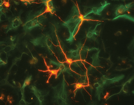 | beta ‑III Tubulin in Rat Cortical Neurons and GFAP in Rat Astrocytes. beta ‑III Tubulin was detected in rat cortical neurons using 5 µg/mL neuron-specific Mouse Anti-Neuron-specific beta ‑III Tubulin Monoclonal Antibody (Catalog # MAB1195). GFAP was detected in rat astrocytes using 10 µg/mL Human GFAP Antigen Affinity-purified Polyclonal Antibody (Catalog # AF2594). Cells were incubated with primary antibodies for 3 hours at room temperature. Cells were stained forbeta‑III Tubulin using the NorthernLights™ 557-conjugated Anti-Mouse IgG Secondary Antibody (red; Catalog # NL007) and for GFAP using the NorthernLights 493-conjugated Anti-Sheep IgG Secondary Antibody (green; Catalog # NL012). View our protocol for Fluorescent ICC Staining of Cells on Coverslips. |
| Immunocytochemistry | |
 | beta ‑III Tubulin and Nestin in Rat Cortical Stem Cells. beta ‑III Tubulin and Nestin were detected in rat cortical stem cells (Catalog # NSC001) using 5 µg/mL neuron-specific Mouse Anti-Neuron-specific beta ‑III Tubulin Monoclonal Antibody (Catalog # MAB1195) and 10 µg/mL Rat Nestin Antigen Affinity-purified Polyclonal Antibody (Catalog # AF2736). Cells were incubated with primary antibodies for 3 hours at room temperature. Cells were stained for beta‑III Tubulin using the NorthernLights™ 557-conjugated Anti-Mouse IgG Secondary Antibody (red; Catalog # NL007) and for Nestin using the NorthernLights 493-conjugated Anti-Goat IgG Secondary Antibody (green; Catalog # NL003). Tissue was counterstained with DAPI (blue). View our protocol for Fluorescent ICC Staining of Cells on Coverslips. |
| Simple Western | |
 | Detection of Rat beta ‑III Tubulin by Simple WesternTM. Simple Western lane view shows lysates of rat cortical neurons, loaded at 0.2 mg/mL. A specific band was detected for beta ‑III Tubulin at approximately 56 kDa (as indicated) using 10 µg/mL of Mouse Anti-Neuron-specific beta ‑III Tubulin Monoclonal Antibody (Catalog # MAB1195). This experiment was conducted under reducing conditions and using the 12-230 kDa separation system. |
カートに商品を
追加しました。
追加しました。
Related Product & Information
| Entrez Gene IDs | 10381 (Human) |
|---|---|
| Background | beta-III Tubulin |
| background_content | Background: beta-III Tubulin beta -III Tubulin, also known as tubulin beta -4, is regarded as a neuron-specific marker. The expression of beta -III Tubulin has been suggested to be one of the earliest markers to signal commitment in primitive neuroepithelium. |
カートに商品を
追加しました。
追加しました。
Citations
R&D Systems personnel manually curate a database that contains references using R&D Systems products.
The data collected includes not only links to publications in PubMed,
but also provides information about sample types, species, and experimental conditions.
- Neurturin Gene Therapy Protects Parasympathetic Function to Prevent Irradiation-Induced Murine Salivary Gland Hypofunction
Authors: JNA Ferreira, C Zheng, IMA Lombaert, CM Goldsmith, AP Cotrim, JM Symonds, VN Patel, MP Hoffman
Mol Ther Methods Clin Dev, 2018;9(0):172-180.
Species: Mouse
Sample Type: Whole Tissue
Application: IHC - Multicolor quantitative confocal imaging cytometry
Authors: DL Coutu, KD Kokkaliari, L Kunz, T Schroeder
Nat. Methods, 2018;15(1):39-46.
Species: Mouse
Sample Type: Whole Tissue
Application: IHC - Uncovering inherent cellular plasticity of multiciliated ependyma leading to ventricular wall transformation and hydrocephalus
Authors: K Abdi, CH Lai, P Paez-Gonza, M Lay, J Pyun, CT Kuo
Nat Commun, 2018;9(1):1655.
Species: Mouse
Sample Type: Whole Tissue
Application: IHC - Generation of multidrug resistant human tissues by overexpression of the ABCG2 multidrug transporter in embryonic stem cells
Authors: Z Erdei, A Schamberge, G Török, K Szebényi, G Várady, TI Orbán, L Homolya, B Sarkadi, Á Apáti
PLoS ONE, 2018;13(4):e0194925.
Species: Human
Sample Type: Whole Cells
Application: ICC - Salivary glands regenerate after radiation injury through SOX2-mediated secretory cell replacement
Authors: E Emmerson, AJ May, L Berthoin, N Cruz-Pache, S Nathan, AJ Mattingly, JL Chang, WR Ryan, AD Tward, SM Knox
EMBO Mol Med, 2018;0(0):.
Species: Human
Sample Type: Whole Tissue
Application: IHC - Paraffin embedded - Nitrosative damage during retrovirus infection-induced neuropathic pain
Authors: P Chauhan, WS Sheng, S Hu, S Prasad, JR Lokensgard
J Neuroinflammation, 2018;15(1):66.
Species: Mouse
Sample Type: Whole Tissue
Application: IHC - Regulation of neuritogenesis in hippocampal neurons using stiffness of extracellular microenvironment
Authors: A Tanaka, Y Fujii, N Kasai, T Okajima, H Nakashima
PLoS ONE, 2018;13(2):e0191928.
Species: Rat
Sample Type: Whole Cells
Application: ICC - Lineage-Specific Differentiation Is Influenced by State of Human Pluripotency
Authors: JH Lee, S Laronde, TJ Collins, Z Shapovalov, B Tanasijevi, JD McNicol, A Fiebig-Com, YD Benoit, JB Lee, RR Mitchell, M Bhatia
Cell Rep, 2017;19(1):20-35.
Species: Human
Sample Type: Whole Cells
Application: ICC - An Activating STAT3 Mutation Causes Neonatal Diabetes through Premature Induction of Pancreatic Differentiation
Authors: J Saarimäki-, D Balboa, MA Russell, J Saarikettu, M Kinnunen, S Keskitalo, A Malhi, C Valensisi, C Andrus, S Eurola, H Grym, J Ustinov, K Wartiovaar, RD Hawkins, O Silvennoin, M Varjosalo, NG Morgan, T Otonkoski
Cell Rep, 2017;19(2):281-294.
Species: Human
Sample Type: Whole Cells
Application: ICC - Transfer of pathogenic and nonpathogenic cytosolic proteins between spinal cord motor neurons in vivo in chimeric mice
Authors: EV Thomas, WA Fenton, J McGrath, AL Horwich
Proc. Natl. Acad. Sci. U.S.A, 2017;0(0):.
Species: Mouse
Sample Type: Whole Tissue
Application: IHC
カートに商品を
追加しました。
追加しました。
製品情報は掲載時点のものですが、価格表内の価格については随時最新のものに更新されます。お問い合わせいただくタイミングにより製品情報・価格などは変更されている場合があります。
表示価格に、消費税等は含まれていません。一部価格が予告なく変更される場合がありますので、あらかじめご了承下さい。