
抗Human DC-SIGN+DC-SIGNR抗体(Anti-Human DC-SIGN+DC-SIGNR antibody)
掲載日情報:2018/11/26 現在Webページ番号:29240

Human DC-SIGN+DC-SIGNRに対する抗体(Anti-Human DC-SIGN+DC-SIGNR )です。
※ 本製品は研究用です。研究用以外には使用できません。
追加しました。
価格
[在庫・価格 :2024年05月01日 20時55分現在]
| 詳細 | 商品名 |
|
文献数 | ||
|---|---|---|---|---|---|
|
Anti-Human DC-SIGN+DC-SIGNR MAb (Clone 120612) |
|
1 | |||
|
Anti-Human DC-SIGN+DC-SIGNR MAb (Clone 120612) |
|
1 | |||
|
Anti-Human DC-SIGN+DC-SIGNR MAb (Clone 120612) |
|
1 | |||
[在庫・価格 :2024年05月01日 20時55分現在]
Anti-Human DC-SIGN+DC-SIGNR MAb (Clone 120612)
文献数: 1
- 商品コード:MAB1621-100
- メーカー:RSD
- 包装:100μg
- 価格:¥101,000
- 在庫:無(未発注)
- 納期:10日程度 ※※ 表示されている納期は弊社に在庫がなく、取り寄せた場合の目安納期となります。
- 法規制等:
Anti-Human DC-SIGN+DC-SIGNR MAb (Clone 120612)
文献数: 1
- 商品コード:MAB1621-500
- メーカー:RSD
- 包装:500μg
- 価格:¥232,000
- 在庫:無(未発注)
- 納期:10日程度 ※※ 表示されている納期は弊社に在庫がなく、取り寄せた場合の目安納期となります。
- 法規制等:
Anti-Human DC-SIGN+DC-SIGNR MAb (Clone 120612)
文献数: 1
- 商品コード:MAB1621-SP
- メーカー:RSD
- 包装:25μg
- 価格:¥30,000
- 在庫:無(未発注)
- 納期:2~3週間 ※※ 表示されている納期は弊社に在庫がなく、取り寄せた場合の目安納期となります。
- 法規制等:
追加しました。
Product Details
| Species Reactivity | Human |
|---|---|
| Label | Unconjugated |
| Immunogen | NIH-3T3 mouse embryonic fibroblast cell line transfected with human DC-SIGNRAccession # Q9H2X3 |
| Source | Monoclonal Mouse IgG2A Clone # 120612 |
| Purification | Protein A or G purified from hybridoma culture supernatant |
| Specificity | Recognizes both human DC-SIGN and human DC-SIGNR on transfected cells. Does not react with parental mouse cells or irrelevant transfectants. |
追加しました。
Applications and Data
| Recommended Concentration | Sample | |
| Flow Cytometry | 2.5 µg/106 cells | See below |
| Immunohistochemistry | 8-25 µg/mL | See below |
| CyTOF-ready | Ready to be labeled using established conjugation methods. No BSA or other carrier proteins that could interfere with conjugation. | |
| Flow Cytometry | |
|---|---|
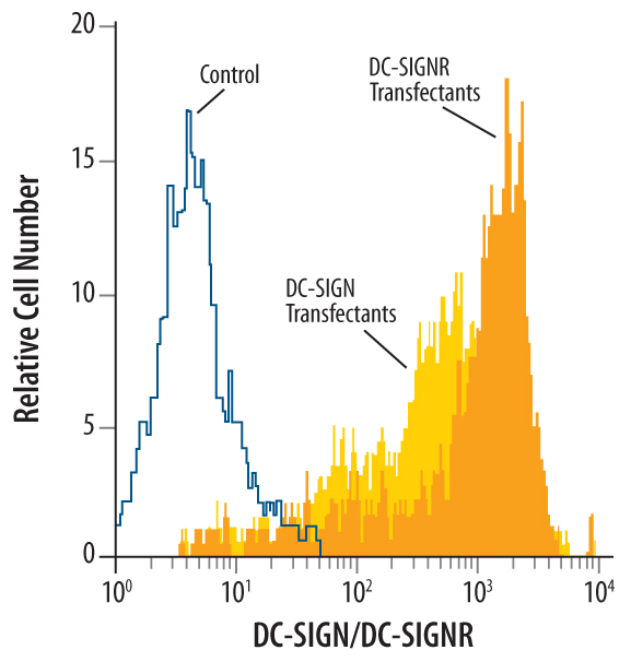 | Detection of DC‑SIGN+DC‑SIGNR in Human DC‑SIGN or DC-SIGNR Transfected 3T3 Mouse Cell Line by Flow Cytometry. Human DC‑SIGN and DC‑SIGNR transfected 3T3 mouse embryonic fibroblast cell line were stained with Mouse Anti-Human DC‑SIGN+DC‑SIGNR Monoclonal Antibody (Catalog # MAB1621, filled histograms) or isotype control antibody (Catalog # MAB003, open histogram), followed by Phycoerythrin-conjugated Anti-Mouse IgG F(ab')2 Secondary Antibody (Catalog # F0102B). |
| Flow Cytometry | |
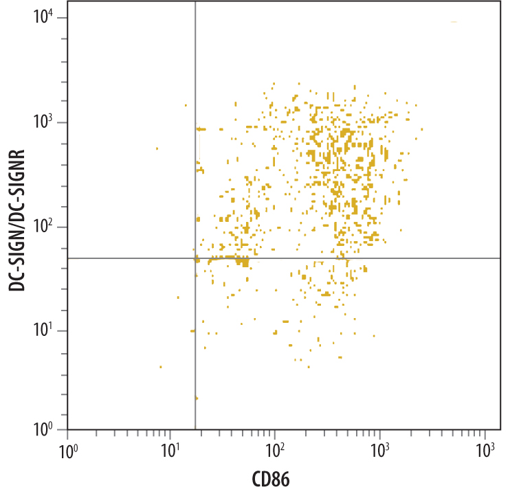 | Detection of DC‑SIGN+DC‑SIGNR in Human Monocyte Derived Dendritic Cells by Flow Cytometry. Human monocyte derived dendritic cells were stained with Mouse Anti-Human DC‑SIGN+ DC‑SIGNR Monoclonal Antibody (Catalog # MAB1621), followed by PE-conjugated anti-mouse secondary antibody (Catalog # F0102B) and Human B7-2/CD86 Fluorescein-conjugated Monoclonal Antibody (Catalog # FAB141F).Quadrant markers were set based on isotype control antibody staining (Catalog # MAB003). |
| Immunohistochemistry | |
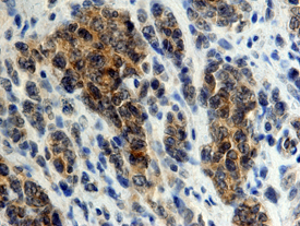 | DC‑SIGN+DC‑SIGNR in Human Lymphoma. DC‑SIGN+DC‑SIGNR was detected in immersion fixed paraffin-embedded sections of human lymphoma using 25 µg/mL Mouse Anti-Human DC‑SIGN+DC‑SIGNR Monoclonal Antibody (Catalog # MAB1621) overnight at 4 °C. Tissue was stained with the Anti-Mouse HRP-DAB Cell & Tissue Staining Kit (brown; Catalog # CTS002) and counterstained with hematoxylin (blue). View our protocol for Chromogenic IHC Staining of Paraffin-embedded Tissue Sections. |
追加しました。
Related Product & Information
| Background | DC-SIGN+DC-SIGNR |
|---|---|
| background_content | Background: DC-SIGN+DC-SIGNR DC-SIGN (Dendritic Cell- Specific ICAM-3 Grabbing Non-Integrin) has been shown to play an important role in regulating dendritic cell (DC) and T cell interactions, including antigen presentation to T cells and enhancement of transinfection of CD4+ T cells by HIV-1 (1, 2). Efforts to identify additional type II membrane proteins resulted in the isolation of a molecule related in sequence to DC-SIGN known as DC-SIGNR (DC-SIGN Related) (3, 4). DC-SIGNR shares 73 - 80% amino acid homology with DC-SIGN and is located on human chromosome 19p13.3. Its structure is similar to DC-SIGN and therefore binds mannose residues in a calcium dependent fashion, including ICAM-3 and HIV-1 gp120 (5). DC-SIGNR, also known as L-SIGN (Liver/Lymph node-Specific ICAM-3-Grabbing Non-integrin) and DC-SIGNR, is polymorphic since allelic variations of the exon 4 encoded sequence have been isolated (5). This is further supported by a study demonstrating the ability to isolate a large repertoire of DC-SIGNR transcripts largely the result of alternative splicing of the 7 coding exons (6). L-SIGN/DC-SIGNR is primarily transcribed in the liver and lymph nodes but not in monocyte derived DC (5). Expression of L-SIGN/DC-SIGNR is restricted to endothelial cells derived from liver sinusoids, lymph nodes sinuses and capillaries (7) although variable expression in placenta and some monocytic cell lines has also been reported, including both membrane and soluble isoforms of the protein (6). Expression of DC-SIGN is induced during the in-vitro generation of DC from either monocytes or bone marrow progenitors, with maximal surface expression at day 7 of culture (1). Immature DC in the skin and mature DC in the tonsil have been demonstrated to express DC-SIGN (8). Analysis of various tissues and cell lines suggests that DC-SIGN expression is restricted to DC (1) although a more recent report finds evidence of expression in placenta, resting monocytes and monocytic cell lines (6). This discrepancy may be partially related to the multiple isoforms of DC-SIGN transcripts, including both membrane and soluble forms, as well as exon splice variants reported in the latter study (6). |
追加しました。
Citations
- Primary Human Placental Trophoblasts are Permissive for Zika Virus (ZIKV) Replication
Authors: KM Aagaard, A Lahon, MA Suter, RP Arya, MD Seferovic, MB Vogt, M Hu, F Stossi, MA Mancini, RA Harris, M Kahr, C Eppes, M Rac, MA Belfort, CS Park, D Lacorazza, R Rico-Hesse
Sci Rep, 2017;7(0):41389.
Species: Human
Sample Type: Whole Cells
Application: ICC - Uukuniemi virus as a tick-borne virus model
J Virol, 2016;0(0):.
Species: Human
Sample Type: Whole Cells
Application: Neut - Binding of HIV-1 gp120 to DC-SIGN promotes ASK-1-dependent activation-induced apoptosis of human dendritic cells.
Authors: Chen Y, Hwang S, Chan V, Chung N, Wang S, Li Z, Ma J, Lin C, Hsieh Y, Chang K, Kung S, Wu Y, Chu C, Tai H, Gao G, Zheng B, Yokoyama K, Austyn J, Lin C
PLoS Pathog, 2013;9(1):e1003100.
Species: Human
Sample Type: Whole Cells
Application: Neut - Vaccine protection by live, attenuated simian immunodeficiency virus in the absence of high-titer antibody responses and high-frequency cellular immune responses measurable in the periphery.
Authors: Mansfield K, Lang SM, Gauduin MC, Sanford HB, Lifson JD, Johnson RP, Desrosiers RC
J. Virol., 2008;82(8):4135-48.
Species: Primate - Macaca mulatta (Rhesus Macaque)
Sample Type: Whole Cells
Application: ICC - SIV-associated myocarditis: viral and cellular correlates of inflammation severity.
Authors: Yearley JH, Pearson C, Carville A, Shannon RP, Mansfield KG
AIDS Res. Hum. Retroviruses, 2006;22(6):529-40.
Species: Primate - Macaca mulatta (Rhesus Macaque)
Sample Type: Whole Tissue
Application: IHC Paraffin-embedded - CCR5-, DC-SIGN-dependent endocytosis and delayed reverse transcription after human immunodeficiency virus type 1 infection in human astrocytes.
Authors: Deiva K, Khiati A, Hery C, Salim H, Leclerc P, Horellou P, Tardieu M
AIDS Res. Hum. Retroviruses, 2006;22(11):1152-61.
Species: Human
Sample Type: Whole Cells
Application: Flow - Rhesus macaque dendritic cells efficiently transmit primate lentiviruses independently of DC-SIGN.
Authors: Wu L, Bashirova AA, Martin TD, Villamide L, Mehlhop E, Chertov AO, Unutmaz D, Pope M, Carrington M, KewalRamani VN
Proc. Natl. Acad. Sci. U.S.A., 2002;99(3):1568-73.
Species: Human
Sample Type: Whole Cells
Application: Flow
追加しました。
製品情報は掲載時点のものですが、価格表内の価格については随時最新のものに更新されます。お問い合わせいただくタイミングにより製品情報・価格などは変更されている場合があります。
表示価格に、消費税等は含まれていません。一部価格が予告なく変更される場合がありますので、あらかじめご了承下さい。