
抗Human Pax6抗体(Anti-Human Pax6 antibody)
掲載日情報:2018/11/26 現在Webページ番号:195157

Human Pax6に対する抗体(Anti-Human Pax6 )です。
※ 本製品は研究用です。研究用以外には使用できません。
カートに商品を
追加しました。
追加しました。
価格
[在庫・価格 :2025年11月03日 13時35分現在]
※ 表示されている納期は弊社に在庫が無く、取り寄せた場合の納期目安となります。
| 詳細 | 商品名 |
|
文献数 | ||
|---|---|---|---|---|---|
|
Anti-Human Pax6 Affinity Purified Polyclonal Ab |
|
6 | |||
|
Anti-Human Pax6 Affinity Purified Polyclonal Ab |
|
6 | |||


[在庫・価格 :2025年11月03日 13時35分現在]
※ 表示されている納期は弊社に在庫が無く、取り寄せた場合の納期目安となります。
Anti-Human Pax6 Affinity Purified Polyclonal Ab
文献数: 6
- 商品コード:AF8150
- メーカー:RSD
- 包装:100μg
- 価格:¥83,000
- 在庫:無(未発注)
- 納期:10日程度 ※※ 表示されている納期は弊社に在庫がなく、取り寄せた場合の目安納期となります。
- 法規制等:
Anti-Human Pax6 Affinity Purified Polyclonal Ab
文献数: 6
- 商品コード:AF8150-SP
- メーカー:RSD
- 包装:25μg
- 価格:¥28,000
- 在庫:1個
- 納期:2~3週間 ※※ 表示されている納期は弊社に在庫がなく、取り寄せた場合の目安納期となります。
- 法規制等:
カートに商品を
追加しました。
追加しました。
Product Details
| Species Reactivity | Human |
|---|---|
| Label | Unconjugated |
| Immunogen | E. coli-derived recombinant human Pax6Met1-Arg272Accession # P26367 |
| Source | Polyclonal Sheep IgG |
| Purification | Antigen Affinity-purified |
| Specificity | Detects human Pax6 in direct ELISAs. In direct ELISAs, less than 5% cross-reactivity with recombinant human (rh) Pax1, rhPax2, rhPax3, rhPax4, rhPax5, and rhPax7 is observed. |
カートに商品を
追加しました。
追加しました。
Applications and Data
| Recommended Concentration | Sample | |
| Western Blot | 0.5 µg/mL | See below |
| Simple Western | 5 µg/mL | See below |
| Immunocytochemistry | 5-15 µg/mL | See below |
| Intracellular Staining by Flow Cytometry | 0.25 µg/106 cells | See below |
| Western Blot | |
|---|---|
 | Detection of Human and Rat Pax6 by Western Blot. Western blot shows lysates of HeLa human cervical epithelial carcinoma cell line and rat cortical stem cells. PVDF membrane was probed with 0.5 µg/mL of Sheep Anti-Human Pax6 Antigen Affinity-purified Polyclonal Antibody (Catalog # AF8150) followed by HRP-conjugated Anti-Sheep IgG Secondary Antibody (Catalog # HAF016). A specific band was detected for Pax6 at approximately 48-50 kDa (as indicated). This experiment was conducted under reducing conditions and using Immunoblot Buffer Group 1. |
| Immunocytochemistry | |
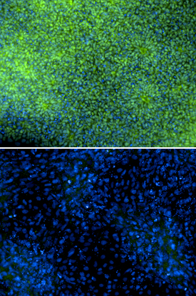 | Pax6 in SA01 Human Embryonic Stem Cells. Immersion fixed SA01 human embryonic stem cells were differentiated for 6 days with Recombinant Human Noggin (Catalog # 6057‑NG) and SB431542 (upper panel) or differentiated for 6 days with Recombinant Human BMP-4 (negative control, lower panel; Catalog # 314-BP). Pax6 was detected using Sheep Anti-Human Pax6 Antigen Affinity-purified Polyclonal Antibody (Catalog # AF8150) at 5 µg/mL. Cells were stained using an Alexa Fluor 488-conjugated Anti-Sheep IgG Secondary Antibody (green) and counterstained with DAPI (blue). Specific staining was localized to nuclei. Images courtesy of Dr. Ron McKay, Leiber Institute for Brain Development, Baltimore, Maryland, USA. |
| Intracellular Staining by Flow Cytometry | |
 | Detection of Pax6 in HeLa Human Cell Line by Flow Cytometry. HeLa human cervical epithelial carcinoma cell line was stained with Sheep Anti-Human Pax6 Affinity-Purified Polyclonal Antibody (Catalog # AF8150, filled histogram) or Sheep IgG control Antibody (Catalog # 5-001-A, open histogram) followed by anti-Sheep IgG PE-conjugated Secondary Antibody (Catalog # F0126). To facilitate intracellular staining, cells were fixed and permeabilized with FlowX FoxP3 Fixation & Permeabilization Buffer Kit (Catalog # FC012). View our protocol for Staining Intracellular Molecules. |
| Simple Western | |
 | Detection of Human Pax6 by Simple WesternTM. Simple Western lane view shows lysates of HeLa human cervical epithelial carcinoma cell line, loaded at 0.2 mg/mL. A specific band was detected for Pax6 at approximately 59 kDa (as indicated) using 5 µg/mL of Sheep Anti-Human Pax6 Antigen Affinity-purified Polyclonal Antibody (Catalog # AF8150) followed by 1:50 dilution of HRP-conjugated Anti-Sheep IgG Secondary Antibody (Catalog # HAF016). This experiment was conducted under reducing conditions and using the12-230 kDa separation system. |
カートに商品を
追加しました。
追加しました。
Related Product & Information
| Long Name | Paired Box Gene 6 |
|---|---|
| Background | Pax6 |
| background_content | Background: Pax6 Pax6 (paired box 6; also Oculorhombin) is a 48‑50 kDa member of the paired homeobox family of transcription factors. It is expressed in developing optic vesicle, olfactory dopaminergic neurons, and pancreatic endocrine cells. Pax6 is a transactivating protein that interacts with MAF, CDX2 and SOX2. Human Pax6 is 422 amino acids (aa) in length. It contains an N-terminal paired box DNA-binding domain (aa 4‑130), a Gly-rich central region (aa 131‑209), a homeodomain (aa 213‑269) and a C-terminal Pro/Ser/Thr-rich regulatory domain (aa 279‑422). Phosphorylation of the C‑terminal domain at Thr281/304/373 promotes Pax6 activity. Multiple splice forms of Pax6 exist. There are alternative start sites at Met137 and a position 34 aa upstream of the standard site. There is also a deletion of aa 22‑26 and 37‑39, plus a 14 aa insertion after Gln47 that generates a C‑terminal DNA binding site. Human and mouse Pax6 are absolutely identical in aa sequence. |
カートに商品を
追加しました。
追加しました。
Citations
R&D Systems personnel manually curate a database that contains references using R&D Systems products.
The data collected includes not only links to publications in PubMed,
but also provides information about sample types, species, and experimental conditions.
- TFAP2C regulates transcription in human naive pluripotency by opening enhancers
Authors: WA Pastor, W Liu, D Chen, J Ho, R Kim, TJ Hunt, A Lukianchik, X Liu, JM Polo, SE Jacobsen, AT Clark
Nat. Cell Biol., 2018;20(5):553-564.
Species: Human
Sample Type: Cell Lysates
Application: WB
カートに商品を
追加しました。
追加しました。
製品情報は掲載時点のものですが、価格表内の価格については随時最新のものに更新されます。お問い合わせいただくタイミングにより製品情報・価格などは変更されている場合があります。
表示価格に、消費税等は含まれていません。一部価格が予告なく変更される場合がありますので、あらかじめご了承下さい。