
抗Human CD7抗体(Anti-Human CD7 antibody)
掲載日情報:2018/11/26 現在Webページ番号:195070

Human CD7に対する抗体(Anti-Human CD7 )です。
※ 本製品は研究用です。研究用以外には使用できません。
カートに商品を
追加しました。
追加しました。
価格
[在庫・価格 :2024年05月18日 00時00分現在]
※ 表示されている納期は弊社に在庫が無く、取り寄せた場合の納期目安となります。
| 詳細 | 商品名 |
|
文献数 | ||
|---|---|---|---|---|---|
|
Anti-Human CD7 Affinity Purified Polyclonal Ab |
|
1 | |||
|
Anti-Human CD7 Affinity Purified Polyclonal Ab |
|
0 | |||
[在庫・価格 :2024年05月18日 00時00分現在]
※ 表示されている納期は弊社に在庫が無く、取り寄せた場合の納期目安となります。
Anti-Human CD7 Affinity Purified Polyclonal Ab
文献数: 1
- 商品コード:AF7579
- メーカー:RSD
- 包装:100μg
- 価格:¥100,000
- 在庫:無(未発注)
- 納期:10日程度 ※※ 表示されている納期は弊社に在庫がなく、取り寄せた場合の目安納期となります。
- 法規制等:
Anti-Human CD7 Affinity Purified Polyclonal Ab
文献数: 0
- 商品コード:AF7579-SP
- メーカー:RSD
- 包装:25μg
- 価格:¥30,000
- 在庫:無(未発注)
- 納期:2~3週間 ※※ 表示されている納期は弊社に在庫がなく、取り寄せた場合の目安納期となります。
- 法規制等:
カートに商品を
追加しました。
追加しました。
Product Details
| Species Reactivity | Human |
|---|---|
| Label | Unconjugated |
| Immunogen | Kidney embryonic cell line HEK293-derived recombinant human CD7Ala26-Pro180Accession # P09564 |
| Source | Polyclonal Sheep IgG |
| Purification | Antigen Affinity-purified |
| Specificity | Detects human CD7 in direct ELISAs and Western blots. In direct ELISAs, less than 1% cross-reactivity with recombinant mouse CD7 is observed. |
カートに商品を
追加しました。
追加しました。
Applications and Data
| Recommended Concentration | Sample | |
| Western Blot | 0.5 µg/mL | See below |
| Flow Cytometry | 2.5 µg/106 cells | See below |
| Immunohistochemistry | 5-15 µg/mL | See below |
| CyTOF-ready | Ready to be labeled using established conjugation methods. No BSA or other carrier proteins that could interfere with conjugation. | |
| Immunocytochemistry | 5-15 µg/mL | See below |
| Western Blot | |
|---|---|
 | Detection of Human CD7 by Western Blot. Western blot shows lysates of MOLT‑4 human acute lymphoblastic leukemia cell line, HepG2 human hepatocellular carcinoma cell line, and human peripheral blood lymphocytes (PBL). PVDF membrane was probed with 0.5 µg/mL of Sheep Anti-Human CD7 Antigen Affinity-purified Polyclonal Antibody (Catalog # AF7579) followed by HRP-conjugated Anti-Sheep IgG Secondary Antibody (Catalog # HAF016). A specific band was detected for CD7 at approximately 35-40 kDa (as indicated). This experiment was conducted under reducing conditions and using Immunoblot Buffer Group 1. |
| Flow Cytometry | |
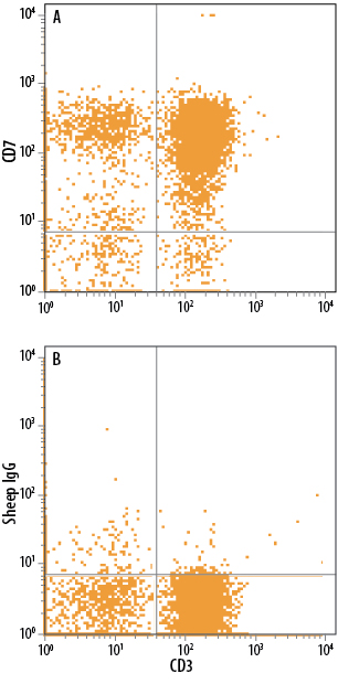 | Detection of CD7 in Human PBMCs by Flow Cytometry. Human peripheral blood mononuclear cells (PBMCs) were stained with Mouse Anti-Human CD3 epsilon APC-conjugated Monoclonal Antibody (Catalog # FAB100A) and either (A) Sheep Anti-Human CD7 Antigen Affinity-purified Polyclonal Antibody (Catalog # AF7579) or (B) Sheep IgG Control (Catalog # 5-001-A) followed by Phycoerythrin-conjugated Anti-Sheep IgG Secondary Antibody (Catalog # F0126). |
| Immunocytochemistry | |
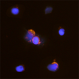 | CD7 in Human PBMCs. CD7 was detected in immersion fixed human peripheral blood mononuclear cells (PBMCs) using Sheep Anti-Human CD7 Antigen Affinity-purified Polyclonal Antibody (Catalog # AF7579) at 15 µg/mL for 3 hours at room temperature. Cells were stained using the NorthernLights™ 557-conjugated Anti-Sheep IgG Secondary Antibody (red; Catalog # NL010) and counterstained with DAPI (blue). Specific staining was localized to cytoplasm and plasma membrane. View our protocol for Fluorescent ICC Staining of Cells on Coverslips. |
| Immunohistochemistry | |
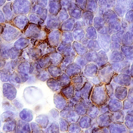 | CD7 in Human Thymus. CD7 was detected in immersion fixed paraffin-embedded sections of human thymus using Sheep Anti-Human CD7 Antigen Affinity-purified Polyclonal Antibody (Catalog # AF7579) at 3 µg/mL overnight at 4 °C. Before incubation with the primary antibody, tissue was subjected to heat-induced epitope retrieval using Antigen Retrieval Reagent-Basic (Catalog # CTS013). Tissue was stained using the Anti-Sheep HRP-DAB Cell & Tissue Staining Kit (brown; Catalog # CTS019) and counterstained with hematoxylin (blue). Specific staining was localized to the plasma membrane. View our protocol for Chromogenic IHC Staining of Paraffin-embedded Tissue Sections. |
カートに商品を
追加しました。
追加しました。
Related Product & Information
| Entrez Gene IDs | 924 (Human); 12516 (Mouse); 303747 (Rat) |
|---|---|
| Background | CD7 |
| background_content | Background: CD7 CD7 (Cluster of Differentiation Antigen 7; also Leu-9, TP41 and GP40) is a 40-44 kDa member of the Ig-superfamily of proteins. It shows restricted expression, being found on fetal thymocytes, CD34+ myeloid and lymphoid progenitor cells, memory CLA- CD45RA+ T cells, and CD56+ IFN-gamma secreting NK cells. CD7 binds to both SECTM1/K12 and galectin-1, and when bound to the latter, initiates complex formation with CD43 in cis. Activation of CD7 may result in either cell proliferation or apoptosis, suggesting a context-dependent signaling mechanism. Mature human CD7 is a 215 amino acid (aa) type I transmembrane glycoprotein. It contains a 155 aa extracellular region (aa 26-180) that shows one V-type Ig-like domain (aa 26-130), and a 39 aa C-terminal cytoplasmic domain. There is one potential alternative splice variant that contains a 79 aa substitution for aa 133-240. Over aa 26-180, human CD7 shares only 43% aa sequence identity with mouse CD7. |
カートに商品を
追加しました。
追加しました。
製品情報は掲載時点のものですが、価格表内の価格については随時最新のものに更新されます。お問い合わせいただくタイミングにより製品情報・価格などは変更されている場合があります。
表示価格に、消費税等は含まれていません。一部価格が予告なく変更される場合がありますので、あらかじめご了承下さい。