
抗Human S100A9抗体(Anti-Human S100A9 antibody)
掲載日情報:2018/11/26 現在Webページ番号:194696

Human S100A9に対する抗体(Anti-Human S100A9 )です。
※ 本製品は研究用です。研究用以外には使用できません。
カートに商品を
追加しました。
追加しました。
価格
[在庫・価格 :2024年05月18日 00時00分現在]
※ 表示されている納期は弊社に在庫が無く、取り寄せた場合の納期目安となります。
| 詳細 | 商品名 |
|
文献数 | ||
|---|---|---|---|---|---|
|
Anti-Human S100A9 Affinity Purified Polyclonal Ab |
|
0 | |||
|
Anti-Human S100A9 Affinity Purified Polyclonal Ab |
|
0 | |||
|
|
|||||
[在庫・価格 :2024年05月18日 00時00分現在]
※ 表示されている納期は弊社に在庫が無く、取り寄せた場合の納期目安となります。
Anti-Human S100A9 Affinity Purified Polyclonal Ab
文献数: 0
- 商品コード:AF5578
- メーカー:RSD
- 包装:100μg
- 価格:¥104,000
- 在庫:無(未発注)
- 納期:10日程度 ※※ 表示されている納期は弊社に在庫がなく、取り寄せた場合の目安納期となります。
- 法規制等:
Anti-Human S100A9 Affinity Purified Polyclonal Ab
文献数: 0
- 商品コード:AF5578-SP
- メーカー:RSD
- 包装:25μg
- 価格:¥30,000
- 在庫:無(未発注)
- 納期:2~3週間 ※※ 表示されている納期は弊社に在庫がなく、取り寄せた場合の目安納期となります。
- 法規制等:
カートに商品を
追加しました。
追加しました。
Product Details
| Species Reactivity | Human |
|---|---|
| Label | Unconjugated |
| Immunogen | E. coli-derived recombinant human S100A9The2-Pro114Accession # P06702 |
| Source | Polyclonal Sheep IgG |
| Purification | Antigen Affinity-purified |
| Specificity | Detects human S100A9 in direct ELISAs and Western blots. In direct ELISAs, less than 5% cross-reactivity with recombinant mouse S100A9 is observed. |
カートに商品を
追加しました。
追加しました。
Applications and Data
| Recommended Concentration | Sample | |
| Western Blot | 0.2 µg/mL | See below |
| Immunohistochemistry | 5-15 µg/mL | See below |
| Immunocytochemistry | 1-25 µg/mL | See below |
| Intracellular Staining by Flow Cytometry | 0.25 µg/106 cells | See below |
| Knockout Validated | S100A9 is specifically detected in MDA‑MB‑468 human breast cancer parental cell line but is not detectable in S100A9 knockout MDA‑MB‑468 cell line. | |
| Western Blot | |
|---|---|
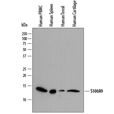 | Detection of Human S100A9 by Western Blot. Western blot shows lysates of human peripheral blood mononuclear cells (PBMC), human spleen tissue, human tonsil tissue, and human cartilage tissue. PVDF membrane was probed with 0.2 µg/mL of Sheep Anti-Human S100A9 Antigen Affinity-purified Polyclonal Antibody (Catalog # AF5578) followed by HRP-conjugated Anti-Sheep IgG Secondary Antibody (Catalog # HAF016). A specific band was detected for S100A9 at approximately 14 kDa (as indicated). This experiment was conducted under reducing conditions and using Immunoblot Buffer Group 1. |
| Immunocytochemistry | |
 | S100A9 in MDA‑MB‑468 Human Cell Line. S100A9 was detected in immersion fixed MDA‑MB‑468 human breast cancer cell line using Sheep Anti-Human S100A9 Antigen Affinity-purified Polyclonal Antibody (Catalog # AF5578) at 1.7 µg/mL for 3 hours at room temperature. Cells were stained using the NorthernLights™ 557-conjugated Anti-Sheep IgG Secondary Antibody (red; Catalog # NL010) and counterstained with DAPI (blue). Specific staining was localized to cytoplasm. View our protocol for Fluorescent ICC Staining of Cells on Coverslips. |
| Immunohistochemistry | |
 | S100A9 in Human Cartilage. S100A9 was detected in immersion fixed paraffin-embedded sections of human cartilage using Sheep Anti-Human S100A9 Antigen Affinity-purified Polyclonal Antibody (Catalog # AF5578) at 1 µg/mL overnight at 4 °C. Tissue was stained using the Anti-Sheep HRP-DAB Cell & Tissue Staining Kit (brown; Catalog # CTS019) and counterstained with hematoxylin (blue). Specific staining was localized to cytoplasm of chondrocytes. View our protocol for Chromogenic IHC Staining of Paraffin-embedded Tissue Sections. |
| Intracellular Staining by Flow Cytometry | |
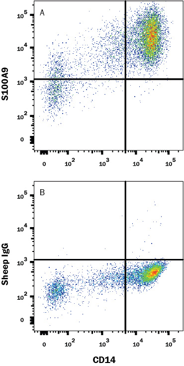 | Detection of S100A9 in Human PBMCs by Flow Cytometry. Human peripheral blood monocytes (PBMC) were stained with Mouse Anti-Human CD14 PE-conjugated Monoclonal Antibody (Catalog # FAB3832P) and either (A) Sheep Anti-Human S100A9 Polyclonal Antibody (Catalog # AF5578) or (B) Sheep IgG Isotype Control (Catalog # 5-001-A) followed by anti-Sheep IgG APC-conjugated Secondary Antibody (Catalog # F0127). To facilitate intracellular staining, cells were fixed with Flow Cytometry Fixation Buffer (Catalog # FC004) and permeabilized with Flow Cytometry Permeabilization/Wash Buffer (Catalog # FC005). View our protocol for Staining Membrane-associated Proteins. |
| Knockout Validated | |
 | Western Blot Shows Human S100A9 Specificity by Using Knockout Cell Line. Western blot shows lysates of MDA‑MB‑468 human breast cancer parental cell line and S100A9 knock out MDA‑MB‑468 cell line (KO). PVDF membrane was probed with 0.2 µg/mL of Sheep Anti-Human S100A9 Antigen Affinity-purified Polyclonal Antibody (Catalog # AF5578) followed by HRP-conjugated Anti-Sheep IgG Secondary Antibody (Catalog # HAF016). A specific band was detected for S100A9 at approximately 14 kDa (as indicated) in the parental MDA‑MB‑468 cell line, but is not detectable in knockout MDA‑MB‑468 cell line. This experiment was conducted under reducing conditions and using Immunoblot Buffer Group 1. |
| Knockout Validated | |
 | S100A9 Specificity is Shown by Immunocytochemistry in Knockout Cell Line. S100A9 was detected in immersion fixed MDA‑MB‑468 human breast cancer cell line but is not detected in S100A9 knockout (KO) MDA-MB-468 cell line using Sheep Anti-Human S100A9 Antigen Affinity-purified Polyclonal Antibody (Catalog # AF5578) at 1.7 µg/mL for 3 hours at room temperature. Cells were stained using the NorthernLights™ 557-conjugated Anti-Sheep IgG Secondary Antibody (red; Catalog # NL010) and counterstained with DAPI (blue). Specific staining was localized to cytoplasm. View our protocol for Fluorescent ICC Staining of Cells on Coverslips. |
カートに商品を
追加しました。
追加しました。
Related Product & Information
| Long Name | S100 Calcium Binding Protein A9 |
|---|---|
| Background | S100A9 |
| background_content | Background: S100A9 Human S100A9 (also MRP-14, Calgranulin-B, and p14) is a 14 kDa member of the S100 family of EF-hand calcium-binding proteins. It is 114 amino acids (aa) in length and contains short sequential modules. There is an N-terminal helical region, followed by a calcium-binding EF-hand domain, two more helical regions, a second EF-hand domain, and three additional helical regions. S100A9 will noncovalently heterodimerize with S100A8. In the presence of calcium, this heterodimer will form a heterotetramer. S100A9 is expressed in granulocytes, monocytes, and macrophages during acute and chronic inflammation. Human S100A9 shares 62% and 57% aa identity with rat and mouse S100A9, respectively. |
カートに商品を
追加しました。
追加しました。
製品情報は掲載時点のものですが、価格表内の価格については随時最新のものに更新されます。お問い合わせいただくタイミングにより製品情報・価格などは変更されている場合があります。
表示価格に、消費税等は含まれていません。一部価格が予告なく変更される場合がありますので、あらかじめご了承下さい。