
抗Enolase 2抗体(Anti-Enolase 2, Sheep-Poly antibody)
掲載日情報:2018/11/26 現在Webページ番号:194613

Enolase 2に対する抗体(Anti-Enolase 2, Sheep-Poly )です。
※ 本製品は研究用です。研究用以外には使用できません。
追加しました。
価格
[在庫・価格 :2026年05月26日 00時00分現在]
| 詳細 | 商品名 |
|
文献数 | ||||||||||||||||||||||||||||||||||||||||||||||||||||||||||||||||||||||||||||||||||
|---|---|---|---|---|---|---|---|---|---|---|---|---|---|---|---|---|---|---|---|---|---|---|---|---|---|---|---|---|---|---|---|---|---|---|---|---|---|---|---|---|---|---|---|---|---|---|---|---|---|---|---|---|---|---|---|---|---|---|---|---|---|---|---|---|---|---|---|---|---|---|---|---|---|---|---|---|---|---|---|---|---|---|---|---|---|
|
Anti-Enolase 2, Sheep-Poly |
|
1 | |||||||||||||||||||||||||||||||||||||||||||||||||||||||||||||||||||||||||||||||||||
|
|||||||||||||||||||||||||||||||||||||||||||||||||||||||||||||||||||||||||||||||||||||
|
Anti-Human/Mouse Enolase 2/Neuron-specific Enolase Aff Pur PAb |
|
1 | |||||||||||||||||||||||||||||||||||||||||||||||||||||||||||||||||||||||||||||||||||


[在庫・価格 :2026年05月26日 00時00分現在]
Anti-Enolase 2, Sheep-Poly
文献数: 1
- 商品コード:AF5169
- メーカー:RSD
- 包装:100μg
- 価格:¥83,000
- 在庫:無(未発注)
- 納期:10日程度 ※※ 表示されている納期は弊社に在庫がなく、取り寄せた場合の目安納期となります。
- 法規制等:
| 説明文 | 別名:2-phospho-D-glycerate hydrolyase Genbank No: 2026 Protein Accession No: P09104 |
||||||
|---|---|---|---|---|---|---|---|
| 別包装品 | 別包装品あり | ||||||
| 法規制等 | |||||||
| 保存条件 | -20℃ | 法規備考 | |||||
| 抗原種 | Human/Mouse | 免疫動物 | Sheep | ||||
| 交差性 | Rat | 適用 | IHC,Simple Western,Western Blot | ||||
| 標識 | Unlabeled | 性状 | Antigen Affinity Purified | ||||
| 吸収処理 | クラス | IgG | |||||
| クロナリティ | Polyclonal | フォーマット | |||||
| 掲載カタログ |
|
||||||
| 製品記事 | Quantikine Human Enolase 2 ELISA Kit |
||||||
| 関連記事 | |||||||
Anti-Human/Mouse Enolase 2/Neuron-specific Enolase Aff Pur PAb
文献数: 1
- 商品コード:AF5169-SP
- メーカー:RSD
- 包装:25μg
- 価格:¥27,000
- 在庫:無(未発注)
- 納期:2~3週間 ※※ 表示されている納期は弊社に在庫がなく、取り寄せた場合の目安納期となります。
- 法規制等:
| 説明文 | ※受注発注品。形状:溶液または凍結乾燥 別名:2-phospho-D-glycerate hydrolyase Genbank No: 2026 Protein Accession No: P09104 |
||||||
|---|---|---|---|---|---|---|---|
| 別包装品 | 別包装品あり | ||||||
| 法規制等 | |||||||
| 保存条件 | -20℃ | 法規備考 | |||||
| 抗原種 | 免疫動物 | Sheep | |||||
| 交差性 | Rat | 適用 | IHC,Simple Western,Western Blot | ||||
| 標識 | Unlabeled | 性状 | Antigen Affinity Purified | ||||
| 吸収処理 | クラス | IgG | |||||
| クロナリティ | Polyclonal | フォーマット | |||||
| 掲載カタログ |
|
||||||
| 製品記事 | 使いっきり抗体 |
||||||
| 関連記事 | |||||||
追加しました。
Product Details
| Species Reactivity | Human, Mouse, Rat |
|---|---|
| Label | Unconjugated |
| Immunogen | E. coli-derived recombinant human Enolase 2/Neuron-specific EnolaseMet1-Leu434Accession # P09104 |
| Source | Polyclonal Sheep IgG |
| Purification | Antigen Affinity-purified |
| Specificity | Detects human and mouse Enolase 2/Neuron-specific Enolase in direct ELISAs. Detects human, mouse, and rat Enolase 2/Neuron-specific Enolase in Western blots. |
追加しました。
Applications and Data
| Recommended Concentration | Sample | |
| Western Blot | 0.2-1 µg/mL | See below |
| Simple Western | 10 µg/mL | See below |
| Immunohistochemistry | 5-15 µg/mL | See below |
| Western Blot | |
|---|---|
 | Detection of Human/Mouse Enolase 2 by Western Blot. Western blot shows lysates of human brain, mouse brain tissue, and BG01V human embryonic stem cells. PVDF membrane was probed with 1 µg/mL of Human/Mouse Enolase 2 Antigen Affinity-purified Polyclonal Antibody (Catalog # AF5169) followed by HRP-conjugated Anti-Sheep IgG Secondary Antibody (Catalog # HAF016). A specific band was detected for Enolase 2 at approximately 46 kDa (as indicated). This experiment was conducted under reducing conditions and using Immunoblot Buffer Group 8. |
| Western Blot | |
 | Detection of Rat Enolase 2/Neuron‑specific Enolase by Western Blot. Western blot shows lysates of rat brain tissue, rat cerebellum tissue, and rat olfactroy bulb tissue. PVDF membrane was probed with 0.2 µg/mL of Sheep Anti-Human/Mouse Enolase 2/Neuron‑specific Enolase Antigen Affinity-purified Polyclonal Antibody (Catalog # AF5169) followed by HRP-conjugated Anti-Sheep IgG Secondary Antibody (Catalog # HAF016). A specific band was detected for Enolase 2/Neuron‑specific Enolase at approximately 47 kDa (as indicated). This experiment was conducted under reducing conditions and using Immunoblot Buffer Group 1. |
| Immunohistochemistry | |
 | Enolase 2 in Human Brain. Enolase 2 was detected in immersion fixed paraffin-embedded sections of human brain (cortex) using Human/Mouse Enolase 2 Antigen Affinity-purified Polyclonal Antibody (Catalog # AF5169) at 10 µg/mL overnight at 4 °C. Before incubation with the primary antibody, tissue was subjected to heat-induced epitope retrieval using Antigen Retrieval Reagent-Basic (Catalog # CTS013). Tissue was stained using the Anti-Sheep HRP-DAB Cell & Tissue Staining Kit (brown; Catalog # CTS019) and counterstained with hematoxylin (blue). Specific staining was localized to cytoplasm. View our protocol for Chromogenic IHC Staining of Paraffin-embedded Tissue Sections. |
| Simple Western | |
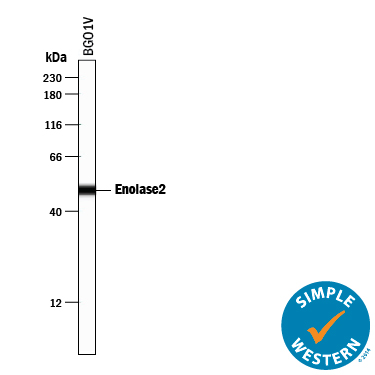 | Detection of Human Enolase 2/Neuron‑specific Enolase by Simple WesternTM. Simple Western lane view shows lysates of BG01V human embryonic stem cells, loaded at 0.2 mg/mL. A specific band was detected for Enolase 2/Neuron‑specific Enolase at approximately 50 kDa (as indicated) using 10 µg/mL of Sheep Anti-Human/Mouse Enolase 2/Neuron‑specific Enolase Antigen Affinity-purified Polyclonal Antibody (Catalog # AF5169) followed by 1:50 dilution of HRP-conjugated Anti-Sheep IgG Secondary Antibody (Catalog # HAF016). This experiment was conducted under reducing conditions and using the 12‑230 kDa separation system. |
追加しました。
Related Product & Information
| Entrez Gene IDs | 2026 (Human); 13807 (Mouse); 24334 (Rat) |
|---|---|
| Background | Enolase 2/Neuron-specific Enolase |
| background_content | Background: Enolase 2/Neuron-specific Enolase Enolase 2 (2-phospho-D-glycerate hydrolyase; also neural enolase and gamma -enolase) is a 46 kDa member of the Enolase family of enzymes. It is expressed in developing neurons and glia, is known to catalyze the generation of phosphoenolpyruvate, and is suggested to possess neurotrophic activity for neurons, likely through an extracellular mechanism. Human Enolase 2 is 434 amino acids (aa) in length. The enzymatic site spans most of the length of the molecule. Enolase 2 exists as both a noncovalently-linked homodimer, or heterodimer with alpha -enolase. Full-length human Enolase 2 is 99% aa identical to both mouse and canine Enolase 2. It shares 83% aa identity with human enolases # 1 and # 3. |
追加しました。
Citations
- Inflammation-induced reversible switch of the neuron-specific enolase promoter from Purkinje neurons to Bergmann glia
Authors: Yusuke Sawada
Sci Rep, 2016;6(0):27758.
Species: Mouse
Sample Type: Whole Tissue
Application: IHC Paraffin-embedded
追加しました。
製品情報は掲載時点のものですが、価格表内の価格については随時最新のものに更新されます。お問い合わせいただくタイミングにより製品情報・価格などは変更されている場合があります。
表示価格に、消費税等は含まれていません。一部価格が予告なく変更される場合がありますので、あらかじめご了承下さい。