
Kidney Cancer Tissue Array, With Matched Adjacent Normal Tissue, 40 Cases/80 Cores|ヒト組織アレイ
掲載日情報:2015/08/07 現在Webページ番号:173348

US Biomax社は、豊富なラインナップの組織アレイ、組織切片(凍結、FFPE)を提供しているメーカーです。
US Biomax社のヒト組織アレイ(Kidney Cancer Tissue Array, With Matched Adjacent Normal Tissue, 40 Cases/80 Cores)をご紹介します。
※ヒト組織アレイは、ドナーからインフォームドコンセントを取得した組織を使用しています。
※ドナーの組織は、HIV、Hepatitis B、Hepatitis Cが陰性であることを確認しています。
※本製品は研究用です。研究用以外には使用できません。
追加しました。
ヒト組織アレイの価格
[在庫・価格 :2026年07月25日 10時35分現在]
| 詳細 | 商品名 |
|
文献数 | ||||||||||||||||||||||||||||||||||||||||||||||||||
|---|---|---|---|---|---|---|---|---|---|---|---|---|---|---|---|---|---|---|---|---|---|---|---|---|---|---|---|---|---|---|---|---|---|---|---|---|---|---|---|---|---|---|---|---|---|---|---|---|---|---|---|---|---|
|
Human, Kidney Cancer with Matched Adjacent Normal Tissue, Tissue Array |
|
本製品は取扱中止になりました | 0 | ||||||||||||||||||||||||||||||||||||||||||||||||||
|
|||||||||||||||||||||||||||||||||||||||||||||||||||||

[在庫・価格 :2026年07月25日 10時35分現在]
Human, Kidney Cancer with Matched Adjacent Normal Tissue, Tissue Array
文献数: 0
- 商品コード:KD801
- メーカー:USB
- 包装:1slide
- 本製品は取扱中止になりました
| 説明文 | |||
|---|---|---|---|
| 法規制等 | |||
| 保存条件 | 法規備考 | ||
| 掲載カタログ |
|
||
| 製品記事 | Cancer Tissue Array TissueArray.Com社 組織アレイ(Tissue array) 選択ガイド |
||
| 関連記事 | TissueArray.Com社 パラフィン組織スライド/組織アレイの仕様変更につきまして |
||
追加しました。
ヒト組織アレイの特長
| Microarray Panel: | Kidney cancer tissue microarray, containing 40 pairs of kidney cancer and matched or unmatched normal adjacent tissue | |
|---|---|---|
| Cores: | 80 | 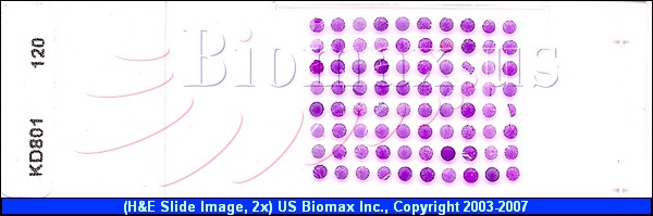 |
| Cases: | 40 | |
| Layout: | 10 cols × 8 rows | |
| Core Diameter: | 1.5 mm | |
| Thickness: | 5 µm | |
| Quality Control: | Anti-Cytokeratin (High MW) confirmed | |
| Applications: | Routine histology procedures including ImmunoHistochemistry (IHC) and In Situ Hybridization (ISH), protocols which can be found on our support page. |
|
| Notes: | Unless specified, all TMA slides are not coated with extra layer of paraffin (tissue cores can be easily seen on the glass), so there is no need to bake, can be directly put into xylene for de-paraffin procedure. | |
*Tissue Microarray Slide Types:
- Unstained: unstained paraffin tissue microarray slide.
- Trial: tissue microarray trial slide. 10% - 25% of cores missing. Good for titrating antibody dilution and experiment conditions. Limited numbers are available, to 2 per item per order. Test slides (tissue arrays with catalog numbers ending with 241 or 242) are recommended as a substitute.
- H and E: Hematoxylin and Eosin stained tissue array slide.
追加しました。
ヒト組織アレイの仕様表
| Pos | No. | Sex | Age | Organ | Pathology diagnosis | TNM | Image | Type † |
|---|---|---|---|---|---|---|---|---|
| A1 | 1 | M | 41 | Kidney | Granular cell carcinoma | T1N0M0 | Image | Malignant |
| A2 | 2 | M | 41 | Kidney | Matched normal cortex of kidney | – | Image | Normal |
| A3 | 3 | M | 65 | Kidney | Clear cell carcinoma | T1N0M0 | Image | Malignant |
| A4 | 4 | M | 65 | Kidney | Matched normal cortex of kidney | – | Image | Normal |
| A5 | 5 | M | 60 | Kidney | Clear cell carcinoma | T2N0M0 | Image | Malignant |
| A6 | 6 | M | 60 | Kidney | Matched normal cortex of kidney | – | Image | Normal |
| A7 | 7 | M | 73 | Kidney | Undifferentiated carcinoma | T1N0M0 | Image | Malignant |
| A8 | 8 | M | 73 | Kidney | Matched normal cortex of kidney and focal lymphocytic cells infiltrated in the stroma | – | Image | Normal |
| A9 | 9 | F | 57 | Kidney | Granular cell carcinoma | T2N0M0 | Image | Malignant |
| A10 | 10 | F | 57 | Kidney | Matched normal cortex of kidney | – | Image | Normal |
| B1 | 11 | M | 67 | Kidney | Clear cell carcinoma | T1N0M0 | Image | Malignant |
| B2 | 12 | M | 67 | Kidney | Matched normal cortex of kidney | – | Image | Normal |
| B3 | 13 | M | 40 | Kidney | Clear cell carcinoma | T1N0M0 | Image | Malignant |
| B4 | 14 | M | 40 | Kidney | Matched normal cortex of kidney | – | Image | Normal |
| B5 | 15 | M | 45 | Kidney | Clear cell carcinoma | T1N0M0 | Image | Malignant |
| B6 | 16 | M | 45 | Kidney | Matched normal cortex of kidney | – | Image | Normal |
| B7 | 17 | F | 49 | Kidney | Clear cell carcinoma | T1N0M0 | Image | Malignant |
| B8 | 18 | F | 49 | Kidney | Matched normal cortex of kidney | – | Image | Normal |
| B9 | 19 | M | 76 | Kidney | Granular-Clear cell carcinoma | T1N0M0 | Image | Malignant |
| B10 | 20 | M | 76 | Kidney | Matched normal cortex of kidney | – | Image | Normal |
| C1 | 21 | M | 46 | Kidney | Clear cell carcinoma | T1N0M0 | Image | Malignant |
| C2 | 22 | M | 46 | Kidney | Matched normal cortex of kidney | – | Image | Normal |
| C3 | 23 | M | 63 | Kidney | Clear cell carcinoma | T1N0M0 | Image | Malignant |
| C4 | 24 | M | 63 | Kidney | Matched normal cortex of kidney | – | Image | Normal |
| C5 | 25 | M | 39 | Kidney | Clear cell carcinoma | T2N0M0 | Image | Malignant |
| C6 | 26 | M | 39 | Kidney | Matched normal cortex of kidney | – | Image | Normal |
| C7 | 27 | M | 67 | Kidney | Clear cell carcinoma | T1N0M0 | Image | Malignant |
| C8 | 28 | M | 67 | Kidney | Matched normal cortex of kidney | – | Image | Normal |
| C9 | 29 | M | 52 | Kidney | Granular cell carcinoma | T2N0M0 | Image | Malignant |
| C10 | 30 | M | 52 | Kidney | Matched normal cortex of kidney | – | Image | Normal |
| D1 | 31 | F | 53 | Kidney | Clear cell carcinoma | T2N0M0 | Image | Malignant |
| D2 | 32 | F | 53 | Kidney | Matched normal cortex of kidney | – | Image | Normal |
| D3 | 33 | M | 26 | Kidney | Clear cell carcinoma | T1N0M0 | Image | Malignant |
| D4 | 34 | M | 26 | Kidney | Matched normal cortex of kidney | – | Image | Normal |
| D5 | 35 | M | 74 | Kidney | Granular cell carcinoma | T1N0M0 | Image | Malignant |
| D6 | 36 | M | 74 | Kidney | Matched normal cortex of kidney | – | Image | Normal |
| D7 | 37 | M | 70 | Kidney | Clear cell carcinoma | T1N0M0 | Image | Malignant |
| D8 | 38 | M | 70 | Kidney | Matched normal cortex of kidney | – | Image | Normal |
| D9 | 39 | F | 68 | Kidney | Clear cell carcinoma | T1N0M0 | Image | Malignant |
| D10 | 40 | F | 68 | Kidney | Medulla of kidney | – | Image | Normal |
| E1 | 41 | F | 56 | Kidney | Clear cell carcinoma | T1N0M0 | Image | Malignant |
| E2 | 42 | F | 56 | Kidney | Matched normal cortex of kidney | – | Image | Normal |
| E3 | 43 | F | 31 | Kidney | Clear cell carcinoma | T2N0M0 | Image | Malignant |
| E4 | 44 | F | 31 | Kidney | Medulla of kidney | – | Image | Normal |
| E5 | 45 | M | 59 | Kidney | Clear cell carcinoma | T1N0M0 | Image | Malignant |
| E6 | 46 | M | 59 | Kidney | Matched normal cortex of kidney | – | Image | Normal |
| E7 | 47 | F | 38 | Kidney | Clear cell carcinoma | T2N0M0 | Image | Malignant |
| E8 | 48 | F | 38 | Kidney | Matched normal cortex of kidney | – | Image | Normal |
| E9 | 49 | M | 56 | Kidney | Granular cell carcinoma | T2N0M0 | Image | Malignant |
| E10 | 50 | M | 56 | Kidney | Matched normal cortex of kidney | – | Image | Normal |
| F1 | 51 | M | 70 | Kidney | Clear cell carcinoma | T1N0M0 | Image | Malignant |
| F2 | 52 | M | 70 | Kidney | Matched normal cortex of kidney | – | Image | Normal |
| F3 | 53 | M | 63 | Kidney | Granular cell carcinoma | T2N0M0 | Image | Malignant |
| F4 | 54 | M | 63 | Kidney | Matched normal cortex of kidney | – | Image | Normal |
| F5 | 55 | M | 47 | Kidney | Clear cell carcinoma | T1N0M0 | Image | Malignant |
| F6 | 56 | M | 47 | Kidney | Matched normal cortex of kidney | – | Image | Normal |
| F7 | 57 | F | 78 | Kidney | Clear cell carcinoma | T1N0M0 | Image | Malignant |
| F8 | 58 | F | 78 | Kidney | Matched normal cortex of kidney | – | Image | Normal |
| F9 | 59 | F | 40 | Kidney | Clear cell carcinoma | T2N0M0 | Image | Malignant |
| F10 | 60 | F | 40 | Kidney | Matched normal cortex of kidney | – | Image | Normal |
| G1 | 61 | M | 58 | Kidney | Clear cell carcinoma | T1N0M0 | Image | Malignant |
| G2 | 62 | M | 58 | Kidney | Medulla of kidney | – | Image | Normal |
| G3 | 63 | M | 49 | Kidney | Granular cell carcinoma | T2N0M0 | Image | Malignant |
| G4 | 64 | M | 49 | Kidney | Matched normal cortex of kidney | – | Image | Normal |
| G5 | 65 | M | 51 | Kidney | Clear cell carcinoma | T2N0M0 | Image | Malignant |
| G6 | 66 | M | 51 | Kidney | Matched normal cortex of kidney | – | Image | Normal |
| G7 | 67 | F | 48 | Kidney | Granular cell carcinoma | T1N0M0 | Image | Malignant |
| G8 | 68 | F | 48 | Kidney | Matched normal cortex of kidney | – | Image | Normal |
| G9 | 69 | M | 43 | Kidney | Granular cell carcinoma | T1N0M0 | Image | Malignant |
| G10 | 70 | M | 43 | Kidney | Matched normal cortex of kidney | – | Image | Normal |
| H1 | 71 | F | 43 | Kidney | Undifferentiated carcinoma | T1N0M0 | Image | Malignant |
| H2 | 72 | F | 43 | Kidney | Matched normal cortex of kidney | – | Image | Normal |
| H3 | 73 | F | 53 | Kidney | Clear cell carcinoma with necrosis | T2N0M0 | Image | Malignant |
| H4 | 74 | F | 53 | Kidney | Medulla of kidney | – | Image | Normal |
| H5 | 75 | M | 54 | Kidney | Clear cell carcinoma | T1N0M0 | Image | Malignant |
| H6 | 76 | M | 54 | Kidney | Matched normal cortex of kidney | – | Image | Normal |
| H7 | 77 | M | 54 | Kidney | Clear cell carcinoma | T2N0M0 | Image | Malignant |
| H8 | 78 | M | 54 | Kidney | Matched normal cortex of kidney | – | Image | Normal |
| H9 | 79 | F | 54 | Kidney | B-cell Lymphoma | T1N0M0 | Image | Malignant |
| H10 | 80 | F | 54 | Kidney | Matched normal cortex of kidney | – | Image | Normal |
*For precise diagnosis, refer to pathology description.
追加しました。
ヒト組織アレイのFAQ
追加しました。
製品情報は掲載時点のものですが、価格表内の価格については随時最新のものに更新されます。お問い合わせいただくタイミングにより製品情報・価格などは変更されている場合があります。
表示価格に、消費税等は含まれていません。一部価格が予告なく変更される場合がありますので、あらかじめご了承下さい。