
抗GFAP抗体 | Anti-GFAP antibody
掲載日情報:2020/04/02 現在Webページ番号:35314

GeneTex社の抗GFAP抗体 | Anti-GFAP antibodyです。
※ 本製品は研究用です。研究用以外には使用できません。
追加しました。
特長
- 高品質の抗体です。
- 幅広い研究分野に関連する抗体を取り揃えています。
- 使用されたアプリケーションや動物種などの情報が充実しています。
- 多数の論文で使用実績がある信頼性の高い抗体です。
追加しました。
価格
[在庫・価格 :2024年04月28日 00時00分現在]
| 詳細 | 商品名 |
|
文献数 | ||||||||||||||||||||||||||||||||||||||||||||||||||||||||||||||||||||||||||||||||||
|---|---|---|---|---|---|---|---|---|---|---|---|---|---|---|---|---|---|---|---|---|---|---|---|---|---|---|---|---|---|---|---|---|---|---|---|---|---|---|---|---|---|---|---|---|---|---|---|---|---|---|---|---|---|---|---|---|---|---|---|---|---|---|---|---|---|---|---|---|---|---|---|---|---|---|---|---|---|---|---|---|---|---|---|---|---|
|
Anti-GFAP, Rabbit-Poly |
|
1 | |||||||||||||||||||||||||||||||||||||||||||||||||||||||||||||||||||||||||||||||||||
|
|||||||||||||||||||||||||||||||||||||||||||||||||||||||||||||||||||||||||||||||||||||
|
Anti-GFAP, Rabbit |
|
1 | |||||||||||||||||||||||||||||||||||||||||||||||||||||||||||||||||||||||||||||||||||
|
|||||||||||||||||||||||||||||||||||||||||||||||||||||||||||||||||||||||||||||||||||||
[在庫・価格 :2024年04月28日 00時00分現在]
Anti-GFAP, Rabbit-Poly
文献数: 1
- 商品コード:GTX100850
- メーカー:GNT
- 包装:100μl
- 価格:¥75,000
- 在庫:1個
- 納期:10日程度 ※※ 表示されている納期は弊社に在庫がなく、取り寄せた場合の目安納期となります。
- 法規制等:
| 説明文 |
別名:glial fibrillary acidic protein,ALXDRD Genbank No: 2670 |
||||||
|---|---|---|---|---|---|---|---|
| 別包装品 | 別包装品あり | ||||||
| 法規制等 | |||||||
| 保存条件 | -20℃ | 法規備考 | |||||
| 抗原種 | Human | 免疫動物 | Rabbit | ||||
| 交差性 | Human/Mouse/Rat | 適用 | IC,IF,IHC,Western Blot | ||||
| 標識 | Unlabeled | 性状 | Purified | ||||
| 吸収処理 | クラス | IgG | |||||
| クロナリティ | Polyclonal | フォーマット | |||||
| 掲載カタログ |
|
||||||
| 製品記事 |
Neuroscience Antibody for Learning and Forming Episodic Memory |
||||||
| 関連記事 |
GeneTex社における抗体の品質管理 |
||||||
Anti-GFAP, Rabbit
文献数: 1
- 商品コード:GTX100850
- メーカー:GNT
- 包装:25μl
- 価格:¥24,000
- 在庫:無(未発注)
- 納期:10日程度 ※※ 表示されている納期は弊社に在庫がなく、取り寄せた場合の目安納期となります。
- 法規制等:
| 説明文 |
別名:glial fibrillary acidic protein,ALXDRD Genbank No: 2670 |
||||||
|---|---|---|---|---|---|---|---|
| 別包装品 | 別包装品あり | ||||||
| 法規制等 | |||||||
| 保存条件 | -20℃ | 法規備考 | |||||
| 抗原種 | Human | 免疫動物 | Rabbit | ||||
| 交差性 | Human/Mouse/Rat | 適用 | IC,IF,IHC,Western Blot | ||||
| 標識 | Unlabeled | 性状 | Purified | ||||
| 吸収処理 | クラス | IgG | |||||
| クロナリティ | Polyclonal | フォーマット | |||||
| 掲載カタログ |
|
||||||
| 製品記事 |
Neuroscience Antibody for Learning and Forming Episodic Memory 使いっきり抗体 |
||||||
| 関連記事 |
GeneTex社における抗体の品質管理 |
||||||
追加しました。
DATA IMAGES
 | GFAP antibody detects GFAP protein at glia cells by immunofluorescent analysis.Sample: DIV9 rat E18 primary cortical neurons were fixed in 4% paraformaldehyde at RT for 15 min.Green: GFAP protein stained by GFAP antibody (GTX100850) diluted at 1:500.Red: beta Tubulin 3/ Tuj1, a neuron cell marker, stained by beta Tubulin 3/ Tuj1 antibody [GT11710] (GTX631836) diluted at 1:500.Blue: Fluoroshield with DAPI (GTX30920). |
 | GFAP antibody detects GFAP protein expression in astrocytes/glia cells on mouse brain by immunohistochemical analysis. Sample: Paraffin-embedded mouse brain. GFAP antibody (GTX100850) diluted at 1:500. Antigen Retrieval: Citrate buffer, pH 6.0, 15 min |
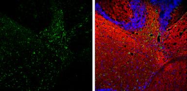 | GFAP antibody detects GFAP protein expression by immunohistochemical analysis.Sample: Frozen-sectioned adult mouse cerebellum. Green: GFAP protein stained by GFAP antibody (GTX100850) diluted at 1:250.Red: beta Tubulin 3/ TUJ1, stained by beta Tubulin 3/ TUJ1 antibody [GT11710] (GTX631836) diluted at 1:500.Blue: Fluoroshield with DAPI (GTX30920). |
 | GFAP antibody detects GFAP protein expression in astrocytes/glia cells on rat brain by immunohistochemical analysis. Sample: Paraffin-embedded rat brain. GFAP antibody (GTX100850) diluted at 1:500. Antigen Retrieval: Citrate buffer, pH 6.0, 15 min |
 | GFAP antibody detects GFAP protein expression by immunohistochemical analysis.Sample: Frozen-sectioned adult mouse hippocampus. Green: GFAP protein stained by GFAP antibody (GTX100850) diluted at 1:250.Red: NeuN, stained by NeuN antibody [2Q158] (GTX30773) diluted at 1:500. |
 | GFAP antibody detects GFAP protein at retinal ganglion cell layer by immunohistochemical analysis.Sample: Frozen sectioned adult mouse retina. Green: GFAP protein stained by GFAP antibody (GTX100850) diluted at 1:250.Red: beta Tubulin 3/ TUJ1, stained by beta Tubulin 3/ TUJ1 antibody [GT11710] (GTX631836) diluted at 1:250.Blue: Fluoroshield with DAPI (GTX30920). |
 | GFAP antibody detects GFAP protein on embryonic mouse brain by immunohistochemical analysis. Sample:Frozen section of embryonic mouse brain (mE18.5). Green: GFAP antibody (GTX100850) diluted at 1:500. Blue: DAPI |
 | GFAP antibodies detects GFAP proteins on embryonic mouse brain by immunohistochemical analysis. Sample: Frozen section of embryonic mouse brain (mE18.5). Green: GFAP antibody (GTX100850) diluted at 1:500. Red: Sox2 antibody [GT1876] (GTX627404) diluted at 1:500. |
 | Rat tissue extract (50 μg) was separated by 10% SDS-PAGE, and the membrane was blotted with GFAP antibody (GTX100850) diluted at 1:10000. |
 | Non-transfected (–) and transfected (+) 293T whole cell extracts (30 μg) were separated by 10% SDS-PAGE, and the membrane was blotted with GFAP antibody (GTX100850) diluted at 1:2500. The HRP-conjugated anti-rabbit IgG antibody (GTX213110-01) was used to detect the primary antibody. |
 | GFAP antibody detects GFAP protein at astrocyte on mouse fore brain by immunohistochemical analysis. Sample: Paraffin-embedded mouse fore brain. GFAP antibody (GTX100850) diluted at 1:500. Antigen Retrieval: Trilogy™ (EDTA based, pH 8.0) buffer, 15min |
 | Mouse tissue extract (50 μg) was separated by 10% SDS-PAGE, and the membrane was blotted with GFAP antibody (GTX100850) diluted at 1:2500. |
 | GFAP antibody detects GFAP protein expression by immunohistochemical analysis.Sample: Frozen-sectioned adult mouse cerebellum. Green: GFAP protein stained by GFAP antibody (GTX100850) diluted at 1:250.Red: beta Tubulin 3/ TUJ1, stained by beta Tubulin 3/ TUJ1 antibody [GT11710] (GTX631836) diluted at 1:500.Blue: Fluoroshield with DAPI (GTX30920). |
 | Various whole cell extracts (30 μg) were separated by 10% SDS-PAGE, and the membrane was blotted with GFAP antibody (GTX100850) diluted at 1:2500. |
追加しました。
製品情報
| Host | Rabbit |
|---|---|
| Clonality | Polyclonal |
| Isotype | IgG |
| Application | WB, ICC/IF, IHC-P, IHC-Fr |
| Reactivity | Human, Mouse, Rat |
追加しました。
APPLICATION
Application Note
*Optimal dilutions/concentrations should be determined by the researcher.| Application | Dilution |
|---|---|
| WB | 1:500-1:3000 |
| ICC/IF | 1:100-1:1000 |
| IHC-P | 1:100-1:1000 |
| IHC-Fr | 1:100-1:1000 |
| Calculated MW | 50 kDa. ( Note ) |
|---|---|
| Positive Control | U87-MG , mouse brain , rat brain , GFAP-transfected 293T |
| Predict Reactivity | Bovine, Pig(>80% identity) |
追加しました。
PROPERTIES
| Form | Liquid |
|---|---|
| Buffer | 1XPBS, 20% Glycerol (pH7). 0.025% ProClin 300 was added as a preservative. |
| Storage | Store as concentrated solution. Centrifuge briefly prior to opening vial. For short-term storage (1-2 weeks), store at 4ºC. For long-term storage, aliquot and store at -20ºC or below. Avoid multiple freeze-thaw cycles. |
| Concentration | 1.72 mg/ml (Please refer to the vial label for the specific concentration.) |
| Antigen Species | Human |
| Immunogen | Recombinant protein encompassing a sequence within the center region of human GFAP. The exact sequence is proprietary. |
| Purification | Purified by antigen-affinity chromatography. |
| Conjugation | Unconjugated |
| Note | For laboratory use only. Not for any clinical, therapeutic, or diagnostic use in humans or animals. Not for animal or human consumption. |
追加しました。
TARGET
| Synonyms | glial fibrillary acidic protein , ALXDRD |
|---|---|
| Cellular Localization | Cytoplasm |
| Background | This gene encodes one of the major intermediate filament proteins of mature astrocytes. It is used as a marker to distinguish astrocytes from other glial cells during development. Mutations in this gene cause Alexander disease, a rare disorder of astrocytes in the central nervous system. Alternative splicing results in multiple transcript variants encoding distinct isoforms. [provided by RefSeq] |
| Database | ・ Gene ID: 2670 GFAP ・ UniProt: P14136 GFAP |
追加しました。
REFERENCEE(抜粋)
| Application Reference | Application/Reactivity |
|---|---|
| Yang YM et al. Acta Radiol 2015; 56 (7) : 837-43 Normalization of T2 relaxation time and apparent diffusion coefficient in relation to the inflammatory changes in the substantia nigra of rats with focal cerebral ischemia. | Application : IHC-P Reactivity : Rat |
追加しました。
製品情報は掲載時点のものですが、価格表内の価格については随時最新のものに更新されます。お問い合わせいただくタイミングにより製品情報・価格などは変更されている場合があります。
表示価格に、消費税等は含まれていません。一部価格が予告なく変更される場合がありますので、あらかじめご了承下さい。