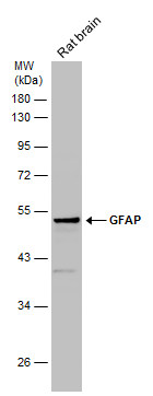
GTX100850 WB Image
Rat tissue extract (50 ug) was separated by 10% SDS-PAGE, and the membrane was blotted with GFAP antibody (GTX100850) diluted at 1:10000.
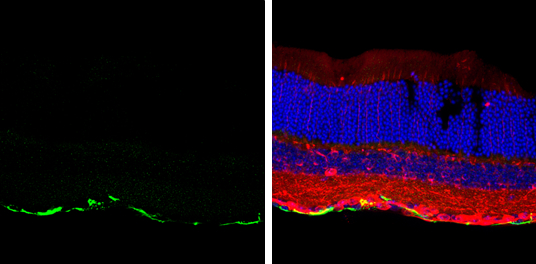
GTX100850 IHC-Fr Image
GFAP antibody detects GFAP protein at retinal ganglion cell layer by immunohistochemical analysis.
Sample: Frozen sectioned adult mouse retina.
Green: GFAP protein stained by GFAP antibody (GTX100850) diluted at 1:250.
Red: beta Tubulin 3/ TUJ1, stained by beta Tubulin 3/ TUJ1 antibody [GT11710] (GTX631836) diluted at 1:250.
Blue: Fluoroshield with DAPI (GTX30920).
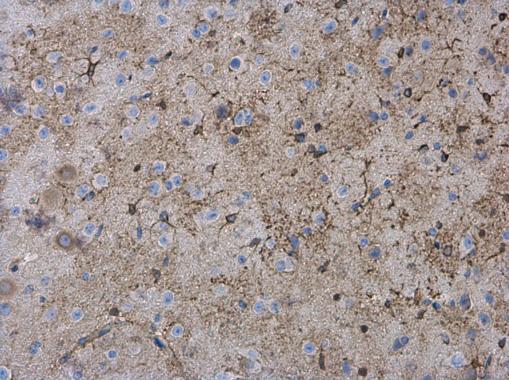
GTX100850 IHC-P Image
GFAP antibody detects GFAP protein expression in astrocytes/glia cells on mouse brain by immunohistochemical analysis.
Sample: Paraffin-embedded mouse brain.
GFAP antibody (GTX100850) diluted at 1:500.
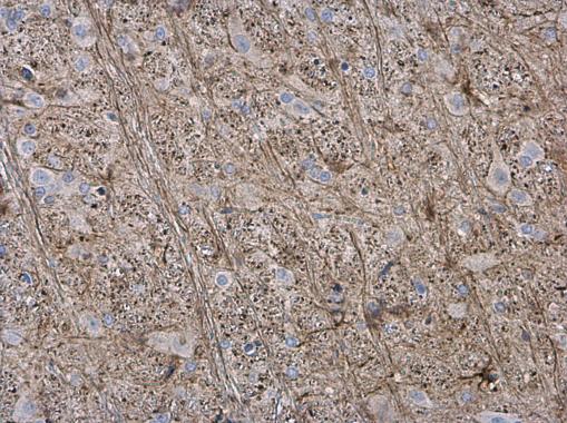
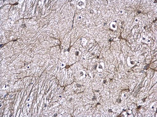
GTX100850 IHC-P Image
GFAP antibody detects GFAP protein expression in astrocytes/glia cells on rat brain by immunohistochemical analysis.
Sample: Paraffin-embedded rat brain.
GFAP antibody (GTX100850) diluted at 1:500.
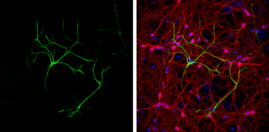
GTX100850 ICC/IF Image
GFAP antibody detects GFAP protein at glia cells by immunofluorescent analysis.
Sample: DIV9 rat E18 primary cortical neurons were fixed in 4% paraformaldehyde at RT for 15 min.
Green: GFAP protein stained by GFAP antibody (GTX100850) diluted at 1:500.
Red: beta Tubulin 3/ Tuj1, a neuron cell marker, stained by beta Tubulin 3/ Tuj1 antibody [GT11710] (GTX631836) diluted at 1:500.
Blue: Fluoroshield with DAPI (GTX30920).
GTX100850 IHC-P Image
GFAP antibody detects GFAP protein at astrocyte on mouse fore brain by immunohistochemical analysis.
Sample: Paraffin-embedded mouse fore brain.
GFAP antibody (GTX100850) diluted at 1:500.
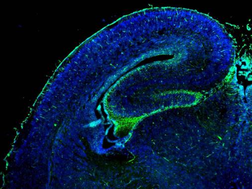
GTX100850 IHC-Fr Image
GFAP antibody detects GFAP protein on embryonic mouse brain by immunohistochemical analysis.
Sample:Frozen section of embryonic mouse brain (mE18.5).
Green: GFAP antibody (GTX100850) diluted at 1:500.
Blue: DAPI
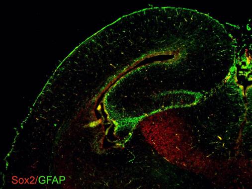
GTX100850 IHC-Fr Image
GFAP antibodies detects GFAP proteins on embryonic mouse brain by immunohistochemical analysis.
Sample: Frozen section of embryonic mouse brain (mE18.5).
Green: GFAP antibody (GTX100850) diluted at 1:500.
Red: Sox2 antibody [GT1876] (GTX627404) diluted at 1:500.
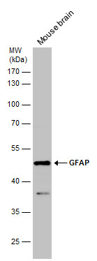
GTX100850 WB Image
Mouse tissue extract (50 ug) was separated by 10% SDS-PAGE, and the membrane was blotted with GFAP antibody (GTX100850) diluted at 1:2500.
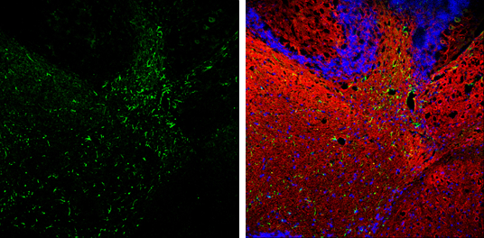
GTX100850 IHC-Fr Image
GFAP antibody detects GFAP protein expression by immunohistochemical analysis.
Sample: Frozen-sectioned adult mouse cerebellum.
Green: GFAP protein stained by GFAP antibody (GTX100850) diluted at 1:250.
Red: beta Tubulin 3/ TUJ1, stained by beta Tubulin 3/ TUJ1 antibody [GT11710] (GTX631836) diluted at 1:500.
Blue: Fluoroshield with DAPI (GTX30920).
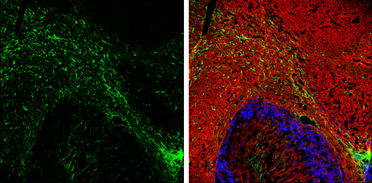
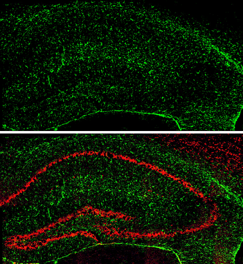
GTX100850 IHC-Fr Image
GFAP antibody detects GFAP protein expression by immunohistochemical analysis.
Sample: Frozen-sectioned adult mouse hippocampus.
Green: GFAP protein stained by GFAP antibody (GTX100850) diluted at 1:250.
Red: NeuN, stained by NeuN antibody [2Q158] (GTX30773) diluted at 1:500.
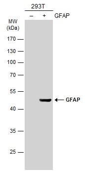
GTX100850 WB Image
Non-transfected (?) and transfected (+) 293T whole cell extracts (30 ug) were separated by 10% SDS-PAGE, and the membrane was blotted with GFAP antibody (GTX100850) diluted at 1:2500. The HRP-conjugated anti-rabbit IgG antibody (GTX213110-01) was used to detect the primary antibody.

GTX100850 WB Image
GFAP antibody detects GFAP protein by western blot analysis.
A. 30 ug U87-MG whole cell extract
B. 30 ug IMR32 whole cell extract
C. 50 ug mouse brain extract
D. 50 ug rat brain extract
10 % SDS-PAGE
GFAP antibody (GTX100850) dilution: 1:2500