
抗ATG5抗体 | Anti-ATG5 Antibody
掲載日情報:2018/07/09 現在Webページ番号:48011

世界最大級の抗体製品数を取り扱うNovus Biologicals社のATG5に対する抗体(anti-ATG5 | antibody ATG5)です。Novus Biologicals社の抗体は数多くの学術論文で使用実績があります。
※本製品は研究用です。研究用以外には使用できません。
価格表左の「文献」アイコンから、使用文献情報一覧が表示できます。

カートに商品を
追加しました。
追加しました。
価格
[在庫・価格 :2024年05月05日 00時00分現在]
※ 表示されている納期は弊社に在庫が無く、取り寄せた場合の納期目安となります。
| 詳細 | 商品名 |
|
文献数 | ||||||||||||||||||||||||||||||||||||||||||||||||||||||||||||||||||||||||||||||||||
|---|---|---|---|---|---|---|---|---|---|---|---|---|---|---|---|---|---|---|---|---|---|---|---|---|---|---|---|---|---|---|---|---|---|---|---|---|---|---|---|---|---|---|---|---|---|---|---|---|---|---|---|---|---|---|---|---|---|---|---|---|---|---|---|---|---|---|---|---|---|---|---|---|---|---|---|---|---|---|---|---|---|---|---|---|---|
|
Anti-ATG5, Rabbit-Poly |
|
194 | |||||||||||||||||||||||||||||||||||||||||||||||||||||||||||||||||||||||||||||||||||
|
|||||||||||||||||||||||||||||||||||||||||||||||||||||||||||||||||||||||||||||||||||||
[在庫・価格 :2024年05月05日 00時00分現在]
※ 表示されている納期は弊社に在庫が無く、取り寄せた場合の納期目安となります。
Anti-ATG5, Rabbit-Poly
文献数: 194
- 商品コード:NB110-53818
- メーカー:NOV
- 包装:0.1ml
- 価格:¥103,000
- 在庫:1個
- 納期:3~4週間 ※※ 表示されている納期は弊社に在庫がなく、取り寄せた場合の目安納期となります。
- 法規制等:

| 説明文 |
レビューあり。Simple Western対応抗体。Keywords:APG5 autophagy 5-like (S. cerevisiae)|APG5LS. cerevisiae)-like|ASPAPG5-LIKE|ATG5 autophagy related 5 homolog (S. cerevisiae)|Autophagy protein 5 Genbank No: 9474 Protein Accession No: Q9H1Y0 |
||||||
|---|---|---|---|---|---|---|---|
| 別包装品 | 別包装品あり | ||||||
| 法規制等 | |||||||
| 保存条件 | -20℃ | 法規備考 | |||||
| 抗原種 | Human | 免疫動物 | Rabbit | ||||
| 交差性 | Alligator/Bovine/Drosophila/Fish/Guinea Pig/Human/Mouse/Porcine/Primate/Rat/Xenopus/Zebrafish | 適用 | ELISA,Electron Microscopy,FCM,IC,IF,IHC,IP,Immunoblotting,Proximity Ligation Assay,RIA,Simple Western,Western Blot | ||||
| 標識 | Unlabeled | 性状 | Antigen Affinity Purified | ||||
| 吸収処理 | クラス | IgG | |||||
| クロナリティ | Polyclonal | フォーマット | |||||
| 掲載カタログ |
|
||||||
| 製品記事 |
抗ATG5抗体 | Anti- ATG5 antibody オートファジーとLC3研究用試薬 |
||||||
| 関連記事 | |||||||
カートに商品を
追加しました。
追加しました。
Image
 | Western Blot: ATG5 Antibody [NB110-53818] - Total protein from Human HeLa and A431 and Mouse MEF cells was separated on a 7.5% gel by SDS-PAGE, transferred to PVDF membrane and blocked in 5% non-fat milk in TBST. The membrane was probed with 2.0 ug/ml anti-ATG5 in 1% non-fat milk in TBST and detected with an anti-rabbit HRP secondary antibody using chemiluminescence. |
 | Immunocytochemistry/Immunofluorescence: ATG5 Antibody [NB110-53818] - HeLa cells were fixed for 10 minutes using 10% formalin and then permeabilized for 5 minutes using 1X PBS + 0.05% Triton-X100. The cells were incubated with anti-ATG5 at 2 ug/ml overnight at 4C and detected with an anti-rabbit Dylight 488 (Green) at a 1:500 dilution. Nuclei were counterstained with DAPI (Blue). Cells were imaged using a 40X objective. |
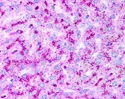 | Immunohistochemistry: ATG5 Antibody [NB110-53818] - Staining of human liver hepatocytes at 2.5ug/ml. 40X magnification. |
 | Western Blot: ATG5 Antibody [NB110-53818] - Analysis in mouse wildtype ES cell lysate (Lane 1) using NB110-53818. Lane 2 is a mouse ATG5 KO ES cell lysate (negative control). Atg5-/- ES cells from Dr. Noboru Mizushima [Mizushima, N. et al. J. Cell Biol. 152 (2001)] Photo courtesy of Dr. Beth Levine, UT Southwestern Medical Center. |
 | Immunocytochemistry/Immunofluorescence: ATG5 Antibody [NB110-53818] - Staining of SY5Y cells at 1:250. Incubated overnight at 4 degrees. Photo courtesy of an anonymous collaborator. |
 | Immunocytochemistry/Immunofluorescence: ATG5 Antibody [NB110-53818] - HeLa cells were fixed for 10 minutes using 10% formalin and then permeabilized for 5 minutes using 1X TBS + 0.5% Triton-X100. The cells were incubated with anti-ATG5 [NB110-53818] at a 1:200 dilution overnight at 4C and detected with an anti-rabbit Dylight 488 (Green) at a 1:500 dilution. Alpha tubulin (DM1A) NB100-690 was used as a co-stain at a 1:1000 dilution and detected with an anti-mouse Dylight 550 (Red) at a 1:500 dilution. Nuclei were counterstained with DAPI (Blue). Cells were imaged using a 40X objective. |
 | Immunohistochemistry: ATG5 Antibody [NB110-53818] - Staining mouse intestine using DAB with hematoxylin counterstain. |
 | Immunohistochemistry-Paraffin: ATG5 Antibody [NB110-53818] - Staining of mouse knee. Image from verified customer review. |
 | Simple Western: ATG5 Antibody [NB110-53818] - Simple Western lane view shows a specific band for ATG5 in 0.5 mg/ml of HeLa lysate. This experiment was performed under reducing conditions using the 12-230 kDa separation system. |
カートに商品を
追加しました。
追加しました。
Background
ATG5 (autophagy protein 5, also known as APG5-like, apoptosis-specific protein, APG5L, and ASP) belongs to ATG5 family and is an essential protein for autophagy. It is expressed ubiquitously and the expression increases dramatically in apoptotic cells. ATG5 is localized in cytoplasm (can colocalize with nonmuscle actin) and is induced upon apoptotic stimuli. ATG5-ATG12 conjugate forms a complex with several units of ATG16 and this complex inhibits ATG5-TECPR1 direct interactions. After conjugating to ATG12, ATG5 associates with the isolation membrane to form autophagosomes and this conjugation reaction is mediated by ubiquitin E1-like enzyme Atg7 as well as the E2-like enzyme Atg10. The conjugate detaches from the membrane immediately before or after the completion of autophagosome formation. ATG5 play an important role in the apoptotic process, within the modified cytoskeleton, and its expression is a relatively late event in apoptotic process, occurring downstream of caspase activity.カートに商品を
追加しました。
追加しました。
製品情報は掲載時点のものですが、価格表内の価格については随時最新のものに更新されます。お問い合わせいただくタイミングにより製品情報・価格などは変更されている場合があります。
表示価格に、消費税等は含まれていません。一部価格が予告なく変更される場合がありますので、あらかじめご了承下さい。