
抗HDAC1抗体 | Anti-HDAC1 antibody
掲載日情報:2020/04/13 現在Webページ番号:35142

GeneTex社の抗HDAC1抗体 | Anti-HDAC1 antibodyです。
※ 本製品は研究用です。研究用以外には使用できません。
GeneTex社 ホームページでユーザーレビューが閲覧できます
GeneTex社では製品を使用したユーザーからのレビューを各国から集め、GeneTex社ホームページで公開しています。各ユーザーの使用アプリケーション、試料の由来動物、希釈倍率、ブロッキング試薬といった実験条件, 実験結果画像など参考となる情報が掲載されています。
⇒抗HDAC1抗体(#GTX100513)のユーザーレビューはこちら。
| Rating |   | ( Average 4 based on 1 users reviews) |
| Application | ||
| Western Blot(WB) | | ( Average 4 based on 1 users reviews) |
追加しました。
特長
- 高品質の抗体です。
- 幅広い研究分野に関連する抗体を取り揃えています。
- 使用されたアプリケーションや動物種などの情報が充実しています。
- 多数の論文で使用実績がある信頼性の高い抗体です。
追加しました。
価格
[在庫・価格 :2024年05月17日 00時01分現在]
| 詳細 | 商品名 |
|
文献数 | ||||||||||||||||||||||||||||||||||||||||||||||||||||||||||||||||||||||||||||||||||
|---|---|---|---|---|---|---|---|---|---|---|---|---|---|---|---|---|---|---|---|---|---|---|---|---|---|---|---|---|---|---|---|---|---|---|---|---|---|---|---|---|---|---|---|---|---|---|---|---|---|---|---|---|---|---|---|---|---|---|---|---|---|---|---|---|---|---|---|---|---|---|---|---|---|---|---|---|---|---|---|---|---|---|---|---|---|
|
Anti-HDAC1, Rabbit-Poly |
|
33 | |||||||||||||||||||||||||||||||||||||||||||||||||||||||||||||||||||||||||||||||||||
|
|||||||||||||||||||||||||||||||||||||||||||||||||||||||||||||||||||||||||||||||||||||
|
Anti-HDAC1, Rabbit-Poly |
|
33 | |||||||||||||||||||||||||||||||||||||||||||||||||||||||||||||||||||||||||||||||||||
|
|||||||||||||||||||||||||||||||||||||||||||||||||||||||||||||||||||||||||||||||||||||
[在庫・価格 :2024年05月17日 00時01分現在]
Anti-HDAC1, Rabbit-Poly
文献数: 33
- 商品コード:GTX100513
- メーカー:GNT
- 包装:100μl
- 価格:¥75,000
- 在庫:1個
- 納期:10日程度 ※※ 表示されている納期は弊社に在庫がなく、取り寄せた場合の目安納期となります。
- 法規制等:

| 説明文 |
レビューあり。KO/KDバリデーション済み抗体。 別名:histone deacetylase 1,GON-10,HD1,KDAC1,RPD3,RPD3L1 Genbank No: 3065 |
||||||
|---|---|---|---|---|---|---|---|
| 別包装品 | 別包装品あり | ||||||
| 法規制等 | |||||||
| 保存条件 | -20℃ | 法規備考 | |||||
| 抗原種 | Human | 免疫動物 | Rabbit | ||||
| 交差性 | Human/Mouse/Rat/Zebrafish | 適用 | ChIP,IC,IF,IHC,IP,Western Blot | ||||
| 標識 | Unlabeled | 性状 | Purified | ||||
| 吸収処理 | クラス | IgG | |||||
| クロナリティ | Polyclonal | フォーマット | |||||
| 掲載カタログ |
|
||||||
| 製品記事 |
GeneTex社 オルガネラマーカー抗体 |
||||||
| 関連記事 |
GeneTex社における抗体の品質管理 |
||||||
Anti-HDAC1, Rabbit-Poly
文献数: 33
- 商品コード:GTX100513
- メーカー:GNT
- 包装:25μl
- 価格:¥24,000
- 在庫:1個
- 納期:10日程度 ※※ 表示されている納期は弊社に在庫がなく、取り寄せた場合の目安納期となります。
- 法規制等:
| 説明文 |
レビューあり。KO/KDバリデーション済み抗体。 別名:histone deacetylase 1,GON-10,HD1,KDAC1,RPD3,RPD3L1 Genbank No: 3065 |
||||||
|---|---|---|---|---|---|---|---|
| 別包装品 | 別包装品あり | ||||||
| 法規制等 | |||||||
| 保存条件 | -20℃ | 法規備考 | |||||
| 抗原種 | Human | 免疫動物 | Rabbit | ||||
| 交差性 | Human/Mouse/Rat/Zebrafish | 適用 | ChIP,IC,IF,IHC,IP,Western Blot | ||||
| 標識 | Unlabeled | 性状 | Purified | ||||
| 吸収処理 | クラス | IgG | |||||
| クロナリティ | Polyclonal | フォーマット | |||||
| 掲載カタログ |
|
||||||
| 製品記事 |
使いっきり抗体 |
||||||
| 関連記事 |
GeneTex社における抗体の品質管理 |
||||||
追加しました。
DATA IMAGES
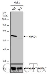 | Wild-type (WT) and HDAC1 knockout (KO) HeLa cell extracts (30 μg) were separated by 10% SDS-PAGE, and the membrane was blotted with HDAC1 antibody (GTX100513) diluted at 1:500. The HRP-conjugated anti-rabbit IgG antibody (GTX213110-01) was used to detect the primary antibody. |
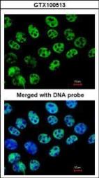
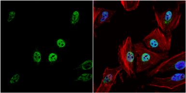 | HDAC1 antibody detects HDAC1 protein at nucleus by immunofluorescent analysis. Sample: HeLa cells were fixed in 4% paraformaldehyde at RT for 15 min. Green: HDAC1 protein stained by HDAC1 antibody (GTX100513) diluted at 1:500. Red: phalloidin, a cytoskeleton marker, stained by phalloidin (invitrogen, A12380) diluted at 1:200. Blue: Hoechst 33342 staining. |
 | Non-transfected (–) and transfected (+) 293T whole cell extracts (30 μg) were separated by 7.5% SDS-PAGE, and the membrane was blotted with HDAC1 antibody (GTX100513) diluted at 1:4000. The HRP-conjugated anti-rabbit IgG antibody (GTX213110-01) was used to detect the primary antibody. |
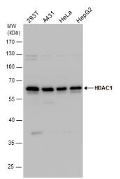 | HDAC1 antibody detects HDAC1 protein by western blot analysis. Various whole cell extracts (30 μg) were separated by 10% SDS-PAGE, and the membrane was blotted with HDAC1 antibody (GTX100513) diluted by 1:1000. The HRP-conjugated anti-rabbit IgG antibody (GTX213110-01) was used to detect the primary antibody. |
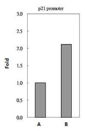 | HDAC1 antibody immunoprecipitates HDAC1 protein-DNA in ChIP experiments. ChIP Sample: 293T whole cell lysate/extract A. 5 μg preimmune rabbit IgG B. 5 μg of HDAC1 antibody (GTX100513) The precipitated DNA was detected by PCR with primer set targeting to p21 promoter. |
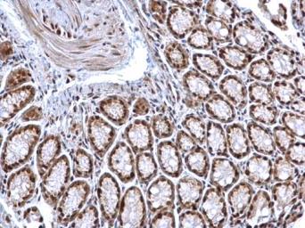 | HDAC1 antibody detects HDAC1 protein at nucleus on mouse colon by immunohistochemical analysis. Sample: Paraffin-embedded mouse colon. HDAC1 antibody (GTX100513) dilution: 1:500. Antigen Retrieval: Trilogy™ (EDTA based, pH 8.0) buffer, 15min |
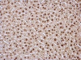 | HDAC1 antibody detects HDAC1 protein at nucleus on mouse liver by immunohistochemical analysis. Sample: Paraffin-embedded mouse liver. HDAC1 antibody (GTX100513) dilution: 1:500. Antigen Retrieval: Trilogy™ (EDTA based, pH 8.0) buffer, 15min |
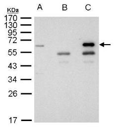 | HDAC1antibody immunoprecipitates HDAC1 protein in IP experiments. IP Sample: 1000 μg 293T whole cell lysate/extract A. 40 μg 293T whole cell lysate/extract B. Control with 2.5 μg of preimmune rabbit IgG C. Immunoprecipitation of HDAC1 protein by 2.5 μg of HDAC1 antibody (GTX100513) 10% SDS-PAGE The immunoprecipitated HDAC1 protein was detected by HDAC1 antibody (GTX100513) diluted at 1:1000. EasyBlot anti-rabbit IgG (GTX221666-01) was used as a secondary reagent. |
 | Immunofluorescence analysis of paraformaldehyde-fixed A431, using HDAC1(GTX100513) antibody at 1:200 dilution. |
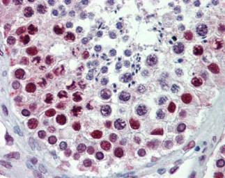 | Immunohistochemical analysis of paraffin-embedded human testis, using HDAC1(GTX100513) antibody(10 μg/ml).
Antigen Retrieval: Trilogy™ (EDTA based, pH 8.0) buffer, 15min |
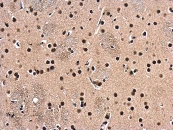 | HDAC1 antibody detects HDAC1 protein at cytoplasm and nucleus in rat brain by immunohistochemical analysis. Sample: Paraffin-embedded rat brain. HDAC1 antibody (GTX100513) diluted at 1:500. Antigen Retrieval: Citrate buffer, pH 6.0, 15 min |
 | HDAC1 antibody detects HDAC1 protein at nucleus in human lung adenocarcinoma by immunohistochemical analysis. Sample: Paraffin-embedded human lung adenocarcinoma. HDAC1 antibody (GTX100513) diluted at 1:250. Antigen Retrieval: Citrate buffer, pH 6.0, 15 min |
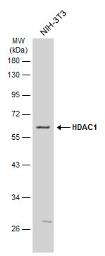 | Whole cell extract (30 μg) was separated by 10% SDS-PAGE, and the membrane was blotted with HDAC1 antibody (GTX100513) diluted at 1:1000. The HRP-conjugated anti-rabbit IgG antibody (GTX213110-01) was used to detect the primary antibody. |
 | Whole cell extract (30 μg) was separated by 10% SDS-PAGE, and the membrane was blotted with HDAC1 antibody (GTX100513) diluted at 1:1000. The HRP-conjugated anti-rabbit IgG antibody (GTX213110-01) was used to detect the primary antibody. |
 | Various whole cell extracts (30 μg) were separated by 10% SDS-PAGE, and the membrane was blotted with HDAC1 antibody (GTX100513) diluted at 1:1000. The HRP-conjugated anti-rabbit IgG antibody (GTX213110-01) was used to detect the primary antibody. |
 | The WB analysis of HDAC1 antibody was published by Brügger V and colleagues in the journal Nat Commun in 2017.PMID: 28139683 |
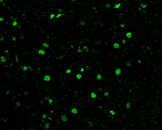 | HDAC1 antibody detects HDAC1 protein at nucleus in rat brain by immunohistochemical analysis. Sample: Paraffin-embedded rat brain. Green: HDAC1 antibody (GTX100513) diluted at 1:200. The signal was developed using goat anti-rabbit IgG antibody (Dylight488) (GTX213110-04). Antigen Retrieval: Citrate buffer, pH 6.0, 15 min |
 | Non-transfected (–) and transfected (+) 293T whole cell extracts (30 μg) were separated by 10% SDS-PAGE, and the membrane was blotted with HDAC1 antibody (GTX100513) diluted at 1:1000. The HRP-conjugated anti-rabbit IgG antibody (GTX213110-01) was used to detect the primary antibody. |
 | The IHC-Fr analysis of HDAC1 antibody was published by Brügger V and colleagues in the journal Nat Commun in 2017.PMID: 28139683 |
追加しました。
製品情報
| Host | Rabbit |
|---|---|
| Clonality | Polyclonal |
| Isotype | IgG |
| Application | WB, ICC/IF, IHC-P, IHC-Fr, IP, ChIP assay |
| Reactivity | Human, Mouse, Rat |
追加しました。
APPLICATION
Application Note
*Optimal dilutions/concentrations should be determined by the researcher.| Application | Dilution |
|---|---|
| WB | 1:500-1:3000 |
| ICC/IF | 1:100-1:1000 |
| IHC-P | 1:100-1:1000 |
| IHC-Fr | Assay dependent |
| IP | 1:100-1:500 |
| ChIP assay | Assay dependent |
| Calculated MW | 55 kDa. ( Note ) |
|---|---|
| Positive Control | 293T , A431 , HeLa , HepG2 , U87-MG , SK-N-SH , NIH3T3 , Rat-2 , *E8.5 embryo |
| Predict Reactivity | Bovine, Xenopus laevis(>80% identity) |
追加しました。
PROPERTIES
| Form | Liquid |
|---|---|
| Buffer | 1XPBS, 20% Glycerol (pH7). 0.01% Thimerosal was added as a preservative. |
| Storage | Store as concentrated solution. Centrifuge briefly prior to opening vial. For short-term storage (1-2 weeks), store at 4ºC. For long-term storage, aliquot and store at -20ºC or below. Avoid multiple freeze-thaw cycles. |
| Concentration | 1 mg/ml (Please refer to the vial label for the specific concentration.) |
| Antigen Species | Human |
| Immunogen | Recombinant protein encompassing a sequence within the center region of human HDAC1. The exact sequence is proprietary. |
| Purification | Purified by antigen-affinity chromatography. |
| Conjugation | Unconjugated |
| Note | For laboratory use only. Not for any clinical, therapeutic, or diagnostic use in humans or animals. Not for animal or human consumption. |
追加しました。
TARGET
| Synonyms | histone deacetylase 1 , GON-10 , HD1 , KDAC1 , RPD3 , RPD3L1 |
|---|---|
| Cellular Localization | Nucleus |
| Background | Histone acetylation and deacetylation, catalyzed by multisubunit complexes, play a key role in the regulation of eukaryotic gene expression. The protein encoded by this gene belongs to the histone deacetylase/acuc/apha family and is a component of the histone deacetylase complex. It also interacts with retinoblastoma tumor-suppressor protein and this complex is a key element in the control of cell proliferation and differentiation. Together with metastasis-associated protein-2, it deacetylates p53 and modulates its effect on cell growth and apoptosis. [provided by RefSeq] |
| Database | ・ Gene ID: 3065 HDAC1 ・ UniProt: Q13547 HDAC1 |
追加しました。
REFERENCE
追加しました。
製品情報は掲載時点のものですが、価格表内の価格については随時最新のものに更新されます。お問い合わせいただくタイミングにより製品情報・価格などは変更されている場合があります。
表示価格に、消費税等は含まれていません。一部価格が予告なく変更される場合がありますので、あらかじめご了承下さい。