
抗ABAT, Mouse-Mono(UMAB179) UltraMAB抗体 | Anti-ABAT, Mouse-Mono(UMAB179) UltraMAB Antibody
掲載日情報:2019/10/29 現在Webページ番号:253517

OriGene社の抗ABAT, Mouse-Mono(UMAB179) UltraMAB抗体です。
本製品は、UltraMAB抗体シリーズ(三次元構造を有する組換え体ヒトタンパク質の全長を抗原として作製したモノクローナル抗体)です。
※本製品は研究用です。研究用以外には使用できません。
カートに商品を
追加しました。
追加しました。
価格
[在庫・価格 :2024年05月18日 09時35分現在]
※ 表示されている納期は弊社に在庫が無く、取り寄せた場合の納期目安となります。
| 詳細 | 商品名 |
|
文献数 | ||||||||||||||||||||||||||||||||||||||||||||||||||||||||||||||||||||||||||
|---|---|---|---|---|---|---|---|---|---|---|---|---|---|---|---|---|---|---|---|---|---|---|---|---|---|---|---|---|---|---|---|---|---|---|---|---|---|---|---|---|---|---|---|---|---|---|---|---|---|---|---|---|---|---|---|---|---|---|---|---|---|---|---|---|---|---|---|---|---|---|---|---|---|---|---|---|---|
|
Anti-ABAT, Mouse-Mono(UMAB179) |
|
0 | |||||||||||||||||||||||||||||||||||||||||||||||||||||||||||||||||||||||||||
|
|||||||||||||||||||||||||||||||||||||||||||||||||||||||||||||||||||||||||||||
[在庫・価格 :2024年05月18日 09時35分現在]
※ 表示されている納期は弊社に在庫が無く、取り寄せた場合の納期目安となります。
Anti-ABAT, Mouse-Mono(UMAB179)
文献数: 0
- 商品コード:UM870071
- メーカー:ORI
- 包装:30μl
- 価格:¥74,000
- 在庫:無(未発注)
- 納期:3~4週間 ※※ 表示されている納期は弊社に在庫がなく、取り寄せた場合の目安納期となります。
- 法規制等:
| 説明文 |
クローン:UMAB179 Gene Accession No: NM_020686 |
||
|---|---|---|---|
| 法規制等 | |||
| 保存条件 | 法規備考 | ||
| 抗原種 | 免疫動物 | Mouse | |
| 交差性 | Human/Mouse/Rat | 適用 | IF,IHC,IP,Western Blot |
| 標識 | Unlabeled | 性状 | |
| 吸収処理 | クラス | IgG | |
| クロナリティ | Monoclonal | フォーマット | |
| 掲載カタログ |
|
||
| 製品記事 |
UltraMAB抗体 |
||
| 関連記事 | |||
カートに商品を
追加しました。
追加しました。
特長
OriGene社は50,000点以上の1次抗体を提供しており、最先端の抗原精製技術により、高品質な抗体をお届けしています。一部抗体製品にはウェスタンブロット用のポジティブコントロールが同梱されます。

OriGene社製品の中でも、特にUltraMAB抗体シリーズは、ヒト組織の免疫染色(IHC)に適した非常に特異性の高いモノクローナル抗体です。抗原にはTrueMAB抗体と同様、組換え体ヒト全長タンパク質を使用しています。1万種類を超えるタンパク質をスポットしたマイクロアレイを用い、標的タンパク質以外の分子との交差反応がないことを確認済みの、癌研究・病理学・解剖学向けの製品です。
カートに商品を
追加しました。
追加しました。
使用例
 | Immunohistochemical staining of paraffin-embedded human liver using ABAT clone UMAB179, mouse monoclonal antibody at 1:400 dilution of 1mg/mL using Polink2 Broad HRP DAB for detection. UM800071 requires heat-induced epitope retrieval with citrate pH6.0 at 110C for 3 min using pressure chamber/cooker. The image shows strong cytoplasmic and membranous staining of the hepatocytes no staining in the bile duct. |
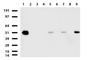 | Western blot of cell lysates (35ug) from 9 different cell lines (1: HepG2, 2: HeLa, 3: SV-T2, 4: A549. 5: COS7, 6: Jurkat, 7: MDCK, 8: PC-12, 9: MCF7). |
 | Western blot analysis of extracts (15ug) from 9 Human tissue by using anti-ABAT monoclonal antibody (1: Testis; 2: Uterus; 3: Breast; 4: Brain; 5: Liver; 6: Ovary; 7: Thyroid gland; 8: colon:;9:Spleen).(1:500) Dilution: 1:500 |
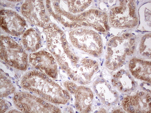 | Immunohistochemical staining of paraffin-embedded human kidney tissue using anti-ABAT mouse monoclonal antibody. Heat-induced epitope retrieval by 1mM EDTA in 10mM Tris buffer (pH8.0) in pressure chamber/cooker at 110C for 3 min, UM800071@ 1:400 shows kidney tubules with strong granular cytoplasmic staining. |
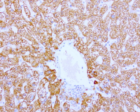
 | Immunohistochemical staining of paraffin-embedded carcinoma of human liver tissue using anti-ABAT mouse monoclonal antibody.(Heat-induced epitope retrieval in pressure chamber/cooker at 110C for 3 min, UM800071(1:400). Images shows cancer cells with strong granular cytoplasmic staining. |
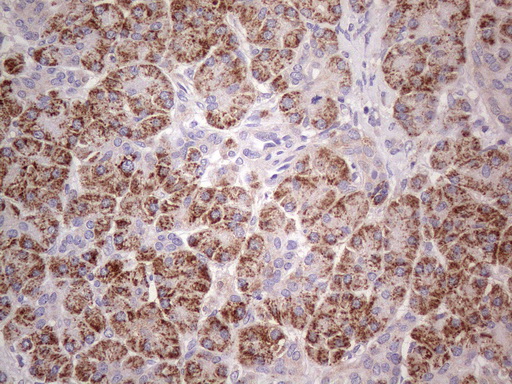 | Immunohistochemical staining of paraffin-embedded human pancreas tissue using anti-ABAT mouse monoclonal antibody. Heat-induced epitope retrieval by 1mM EDTA in 10mM Tris buffer (pH8.0) in pressure chamber/cooker at 110C for 3 min, UM800071(1:400). Images shows exocrine granular cells with strong granular cytoplasmic staining. |
 | Immunohistochemical staining of paraffin-embedded carcinoma of human thyroid tissue using ABAT clone UMAB179, mouse monoclonal antibody. Heat-induced epitope retrieval by 1mM EDTA in 10mM Tris buffer (pH8.0 ) in pressure chamber/cooker at 110C for 3 min, UM800071 was diluted 1:1000 using HRP detection and DAB chromogen. Image shows strong cytoplasmic and membranous staining is present in the tumor cells. |
 | Immunofluorescent staining of HepG2 cells using anti-ABAT mouse monoclonal antibody (UM800071, green, 1:100). Actin filaments were labeled with Alexa Fluor® 594 Phalloidin (red), and nuclear with DAPI (blue). |
 | OriGene overexpression protein microarray chip was immunostained with UltraMAB anti-ABAT mouse monoclonal antibody (UM800071). The positive reactive proteins are highlighted with two red arrows in the enlarged subarray. All the positive controls spotted in this subarray are also labeled for clarification.(1:100) |
カートに商品を
追加しました。
追加しました。
製品情報
| Clone Name | UMAB179 |
|---|---|
| Applications | WB, IHC, IF, 10K-CHIP |
| Recommend Dilution | IHC 1:100~400, |
| Reactivity | Human |
| Host | Mouse |
| Clonality | Monoclonal |
| Immunogen | Human recombinant protein fragment corresponding to amino acids 29-323 of human ABAT(NP_065737) produced in E.coli. |
| Isotype | IgG1 |
| Formulation | PBS (PH 7.3) containing 1% BSA, 50% glycerol and 0.02% sodium azide. |
| Concentration | 0.5~1.0 mg/ml (Lot Dependent) |
| Purification | Purified from cell culture supernatant by affinity chromatography |
| Predicted Protein Size | 53.2 kDa |
| Gene Name | Homo sapiens 4-aminobutyrate aminotransferase (ABAT), transcript variant 1 |
| Background | 4-aminobutyrate aminotransferase (ABAT) is responsible for catabolism of gamma-aminobutyric acid (GABA), an important, mostly inhibitory neurotransmitter in the central nervous system, into succinic semialdehyde. The active enzyme is a homodimer of 50-kD subunits complexed to pyridoxal-5-phosphate. The protein sequence is over 95% similar to the pig protein. GABA is estimated to be present in nearly one-third of human synapses. ABAT in liver and brain is controlled by 2 codominant alleles with a frequency in a Caucasian population of 0.56 and 0.44. The ABAT deficiency phenotype includes psychomotor retardation, hypotonia, hyperreflexia, lethargy, refractory seizures, and EEG abnormalities. Multiple alternatively spliced transcript variants encoding the same protein isoform have been found for this gene. [provided by RefSeq, Jul 2008]. |
| Synonyms | GABA-AT; GABAT; NPD009 |
| Database Link |
カートに商品を
追加しました。
追加しました。
製品情報は掲載時点のものですが、価格表内の価格については随時最新のものに更新されます。お問い合わせいただくタイミングにより製品情報・価格などは変更されている場合があります。
表示価格に、消費税等は含まれていません。一部価格が予告なく変更される場合がありますので、あらかじめご了承下さい。