
抗H3K27me3抗体 | Anti-H3K27me3 Antibody
掲載日情報:2019/11/15 現在Webページ番号:237275

Bioss社の抗H3K27me3抗体(Anti-H3K27me3 Antibody)です。
※ 本製品は研究用です。研究用以外には使用できません。
カートに商品を
追加しました。
追加しました。
Bioss 社抗体製品の特長
Bioss社は、米国ボストンに本拠地を置くメーカーで、約11,000種類の未標識一次抗体と、各抗体に対する最大15種類の標識抗体をそろえています。
- ウエスタンブロット、免疫組織化学(IHC-frozen、IHC-paraffin)、免疫蛍光(IF)、ELISAなどのアプリケーションに適した一次抗体がラインナップされています。
- 一次抗体はすべて、Protein AまたはProtein G精製されています。
- 幅広い研究分野に関連する抗体を取り揃えています。
※最新の適用、交差性情報の詳細は、下記製品情報項の「データシート」リンクをご参照ください。
カートに商品を
追加しました。
追加しました。
価格表
[在庫・価格 :2024年05月18日 00時00分現在]
※ 表示されている納期は弊社に在庫が無く、取り寄せた場合の納期目安となります。
| 詳細 | 商品名 |
|
文献数 | ||
|---|---|---|---|---|---|
|
Anti-H3K27me3 Polyclonal Antibody |
|
0 | |||
[在庫・価格 :2024年05月18日 00時00分現在]
※ 表示されている納期は弊社に在庫が無く、取り寄せた場合の納期目安となります。
Anti-H3K27me3 Polyclonal Antibody
文献数: 0
- 商品コード:bs-53122R
- メーカー:BIS
- 包装:50μg
- 価格:¥80,000
- 在庫:無(未発注)
- 納期:10日程度 ※※ 表示されている納期は弊社に在庫がなく、取り寄せた場合の目安納期となります。
- 法規制等:
カートに商品を
追加しました。
追加しました。
使用例
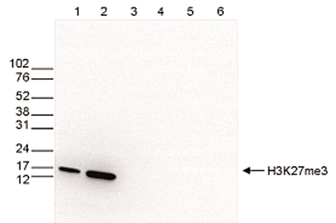 | Western blot was performed on whole cell (25 μg, lane 1) and histone extracts (15 μg, lane 2) from HeLa cells, and on 1 μg of recombinant histone H2A, H2B, H3 and H4 (lane 3, 4, 5 and 6, respectively) using the Bioss antibody against H3K27me3 (Cat. No. bs-53122R) diluted 1:500 in TBS-Tween containing 5% skimmed milk. |
 | A Dot Blot analysis was performed to test the cross-reactivity of the Bioss antibody against H3K27me3 (Cat. No. bs-53122R) with peptides containing other modifications of histone H3 and H4 and the unmodified H3K27 sequence. One hundred to 0.2 pmol of the peptide containing the respective histone modification were spotted on a membrane. The antibody was used at a dilution of 1:5,000. Figure 4 shows a high specificity of the antibody for the modification of interest. Please note that that antibody also recognizes the modification if S28 is phosphorylated. |
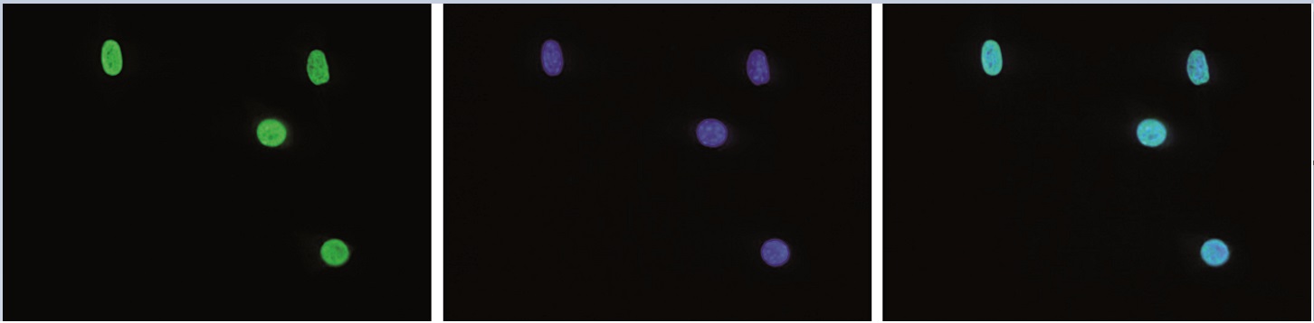 | Mouse NIH3T3 cells were stained with the Bioss antibody against H3K27me3 (Cat. No. bs-53122R) and with DAPI. Cells were fixed with 4% formaldehyde for 10’ and blocked with PBS/TX-100 containing 5% normal goat serum and 1% BSA. The cells were immunofluorescently labeled with the H3K27me3 antibody (left) diluted 1:200 in blocking solution followed by an anti-rabbit antibody conjugated to Alexa488. The middle panel shows staining of the nuclei with DAPI. A merge of the two stainings is shown on the right. |
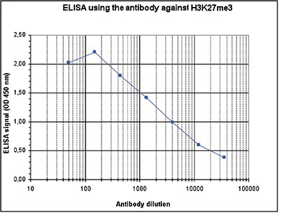 | To determine the titer of the antibody, an ELISA was performed using a serial dilution of H3K27me3 Polyclonal Antibody (Cat. No. bs-53122R). The antigen used was a peptide containing the histone modification of interest. By plotting the absorbance against the antibody dilution, the titer of the antibody was estimated to be 1:3,500. |
 | ChIP assays were performed using human HeLa cells, H3K27me3 Polyclonal Antibody (Cat. No. bs-53122R) and optimized PCR primer sets for qPCR. ChIP was performed with a ChIP-seq kit, using sheared chromatin from 1 million cells. A titration of the antibody consisting of 1, 2, 5, and 10 μg per ChIP experiment was analyzed. IgG (2 μg/IP) was used as negative IP control. QPCR was performed with primers for the promoters of the active genes EIF4A2 and GAPDH as negative controls, and for the coding regions of the inactive genes MYT1 and TSH2B as positive controls. Figure 1 shows the recovery, expressed as a % of input (the relative amount of immunoprecipitated DNA compared to input DNA after qPCR analysis). These results are in accordance with the observation that H3K27me3 is preferably present at inactive genes. |
 | ChIP was performed on sheared chromatin from 1 million HeLaS3 cells using 1 μg of H3K27me3 Polyclonal Antibody (Cat. No. bs-53122R) as described above. The IP’d DNA was subsequently analyzed on an Illumina HiSeq. Library preparation, cluster generation, and sequencing were performed according to the manufacturer’s instructions. The 51 bp tags were aligned to the human genome using the BWA algorithm. The figure shows the enrichment in genomic regions of chromosome 6, surrounding the TSH2B gene (indicated by an arrow; fig A), of chromosome 20, surrounding the MYT1 gene (fig B), and of chromosome 2 and 3 (figure C and D). |
カートに商品を
追加しました。
追加しました。
Background
A core component of nucleosome. Nucleosomes wrap and compact DNA into chromatin, limiting DNA accessibility to the cellular machineries which require DNA as a template. Histones thereby play a central role in transcription regulation, DNA repair, DNA replication and chromosomal stability. DNA accessibility is regulated via a complex set of post-translational modifications of histones, also called histone code, and nucleosome remodeling.カートに商品を
追加しました。
追加しました。
製品情報
| name | H3K27me3 Polyclonal Antibody |
|---|---|
| datasheet | 最新のデータシートはこちらをご確認ください |
| Conjugation | Unconjugated |
| Gene ID | 8350 |
| Swiss Prot | P68431 |
| Source | Polyclonal antibody raised in rabbit against against histone H3, trimethylated at lysine 27 (H3K27me3), using a KLH-conjugated synthetic peptide |
| Purification | Affinity purified polyclonal antibody in PBS containing 0.05% azide and 0.05% ProClin 300. |
カートに商品を
追加しました。
追加しました。
製品情報は掲載時点のものですが、価格表内の価格については随時最新のものに更新されます。お問い合わせいただくタイミングにより製品情報・価格などは変更されている場合があります。
表示価格に、消費税等は含まれていません。一部価格が予告なく変更される場合がありますので、あらかじめご了承下さい。