
抗TP53, Mouse-Mono(UMAB55) UltraMAB抗体 | Anti-TP53, Mouse-Mono(UMAB55) UltraMAB Antibody
掲載日情報:2015/11/06 現在Webページ番号:177921

OriGene社の抗TP53, Mouse-Mono(UMAB55) UltraMAB抗体です。
本製品は、UltraMAB抗体シリーズ(三次元構造を有する組換え体ヒトタンパク質の全長を抗原として作製したモノクローナル抗体)です。
※本製品は研究用です。研究用以外には使用できません。
追加しました。
価格
[在庫・価格 :2024年05月18日 15時35分現在]
| 詳細 | 商品名 |
|
文献数 | ||||||||||||||||||||||||||||||||||||||||||||||||||||||||||||||||||||||||||
|---|---|---|---|---|---|---|---|---|---|---|---|---|---|---|---|---|---|---|---|---|---|---|---|---|---|---|---|---|---|---|---|---|---|---|---|---|---|---|---|---|---|---|---|---|---|---|---|---|---|---|---|---|---|---|---|---|---|---|---|---|---|---|---|---|---|---|---|---|---|---|---|---|---|---|---|---|---|
|
Anti-TP53, Mouse-Mono(UMAB55), UltraMAB |
|
0 | |||||||||||||||||||||||||||||||||||||||||||||||||||||||||||||||||||||||||||
|
|||||||||||||||||||||||||||||||||||||||||||||||||||||||||||||||||||||||||||||
[在庫・価格 :2024年05月18日 15時35分現在]
Anti-TP53, Mouse-Mono(UMAB55), UltraMAB
文献数: 0
- 商品コード:UM570049
- メーカー:ORI
- 包装:30μl
- 価格:¥74,000
- 在庫:無(未発注)
- 納期:3~4週間 ※※ 表示されている納期は弊社に在庫がなく、取り寄せた場合の目安納期となります。
- 法規制等:
| 説明文 |
クローン:UMAB55 Genbank No: 6776 Gene Accession No: NM_000546 |
||
|---|---|---|---|
| 法規制等 | |||
| 保存条件 | 法規備考 | ||
| 抗原種 | 免疫動物 | Mouse | |
| 交差性 | Human | 適用 | IF,IHC,Western Blot |
| 標識 | Unlabeled | 性状 | Affinity Purified |
| 吸収処理 | クラス | IgG | |
| クロナリティ | Monoclonal | フォーマット | |
| 掲載カタログ |
|
||
| 製品記事 |
UltraMAB抗体 |
||
| 関連記事 | |||
追加しました。
UltraMAB抗体について

OriGene社製品の中でも、特にUltraMAB抗体シリーズは、ヒト組織の免疫染色(IHC)に適した非常に特異性の高いモノクローナル抗体です。抗原にはTrueMAB抗体と同様、組換え体ヒト全長タンパク質を使用しています。1万種類を超えるタンパク質をスポットしたマイクロアレイを用い、標的タンパク質以外の分子との交差反応がないことを確認済みの、癌研究・病理学・解剖学向けの製品です。
追加しました。
Product Data
| Immunogen | Full length human recombinant protein of human TP53 (NP_000537) produced in HEK293T cell. | ||
|---|---|---|---|
| Clone Name | Clone UMAB55 | Isotype | IgG1 |
| Species Reactivity | Human | Concentration | 0.5~1.0 mg/ml (Lot Dependent) |
| Guaranteed Application | WB, IHC, IF | Suggested Dilutions | IHC 1:100, |
| Buffer | PBS (PH 7.3) containing 1% BSA, 50% glycerol and 0.02% sodium azide. | ||
| Purification | Purified from mouse ascites fluids by affinity chromatography | ||
追加しました。
Reference Data
| Target Name | Homo sapiens tumor protein p53 (TP53), transcript variant 1 | ||
|---|---|---|---|
| Alternative Name | BCC7; LFS1; P53; TRP53 | ||
| Database Link | NP_000537 | ||
| Function | The protein encoded by this gene is a member of the STAT family of transcription factors. In response to cytokines and growth factors, STAT family members are phosphorylated by the receptor associated kinases, and then form homo- or heterodimers that translocate to the cell nucleus where they act as transcription activators. This protein is activated by, and mediates the responses of many cell ligands, such as IL2, IL3, IL7 GM-CSF, erythropoietin, thrombopoietin, and different growth hormones. Activation of this protein in myeloma and lymphoma associated with a TEL/JAK2 gene fusion is independent of cell stimulus and has been shown to be essential for the tumorigenesis. The mouse counterpart of this gene is found to induce the expression of BCL2L1/BCL-X(L), which suggests the antiapoptotic function of this gene in cells. [provided by RefSeq, Jul 2008]. | ||
| Related Pathway | |||
追加しました。
Image
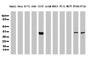
| Western Blot analysis of extracts (35µg) from 11 different cell lines by using anti-TP53 monoclonal antibody (Clone UMAB55) | |
| Immunohistochemical staining of paraffin-embedded Human normal colon tissue and colonrectal cancer tissue using anti-TP53 mouse monoclonal antibody. (UM500049; heat-induced epitope retrieval by 10mM citric buffer, pH6.0, 120C for 3min) | |
| Immunohistochemical staining of paraffin-embedded Human normal lung tissue and lung adenocarcinoma tissue using anti-TP53 mouse monoclonal antibody. (UM500049; heat-induced epitope retrieval by 10mM citric buffer, pH6.0, 120C for 3min) | |
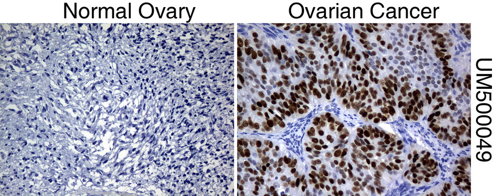
| Immunohistochemical staining of paraffin-embedded Human normal ovary tissue and ovary cancer tissue using anti-TP53 mouse monoclonal antibody. (UM500049; heat-induced epitope retrieval by 10mM citric buffer, pH6.0, 120C for 3min) | |
| Immunohistochemical staining of paraffin-embedded Human normal breast tissue and breast adenocarcinoma tissue using anti-TP53 mouse monoclonal antibody. (UM500049; heat-induced epitope retrieval by 10mM citric buffer, pH6.0, 120C for 3min) | |
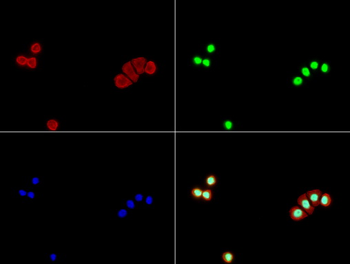
| Immunofluorescent staining of COS7 cells using TP53 mouse monoclonal antibody (UM500049, green). Actin filaments were labeled with TRITC-phalloidin (red), and nuclear with DAPI (blue). The three-color overlay image is located at the bottom-right corner. | |
| Immunofluorescent staining of HT-29 cells using TP53 mouse monoclonal antibody (UM500049, green). Actin filaments were labeled with TRITC-phalloidin (red), and nuclear with DAPI (blue). The three-color overlay image is located at the bottom-right corner. |


追加しました。
製品情報は掲載時点のものですが、価格表内の価格については随時最新のものに更新されます。お問い合わせいただくタイミングにより製品情報・価格などは変更されている場合があります。
表示価格に、消費税等は含まれていません。一部価格が予告なく変更される場合がありますので、あらかじめご了承下さい。