
抗ERCC1, Mouse-Mono(2E12) UltraMAB抗体 | Anti-ERCC1, Mouse-Mono(2E12) UltraMAB Antibody
掲載日情報:2015/11/06 現在Webページ番号:177884

OriGene社の抗ERCC1, Mouse-Mono(2E12) UltraMAB抗体です。
本製品は、UltraMAB抗体シリーズ(三次元構造を有する組換え体ヒトタンパク質の全長を抗原として作製したモノクローナル抗体)です。
※本製品は研究用です。研究用以外には使用できません。
追加しました。
価格
[在庫・価格 :2024年05月18日 12時35分現在]
| 詳細 | 商品名 |
|
文献数 | ||||||||||||||||||||||||||||||||||||||||||||||||||||||||||||||||||||||||||
|---|---|---|---|---|---|---|---|---|---|---|---|---|---|---|---|---|---|---|---|---|---|---|---|---|---|---|---|---|---|---|---|---|---|---|---|---|---|---|---|---|---|---|---|---|---|---|---|---|---|---|---|---|---|---|---|---|---|---|---|---|---|---|---|---|---|---|---|---|---|---|---|---|---|---|---|---|---|
|
Anti-ERCC1, Mouse-Mono(2E12), UltraMAB |
|
0 | |||||||||||||||||||||||||||||||||||||||||||||||||||||||||||||||||||||||||||
|
|||||||||||||||||||||||||||||||||||||||||||||||||||||||||||||||||||||||||||||
[在庫・価格 :2024年05月18日 12時35分現在]
Anti-ERCC1, Mouse-Mono(2E12), UltraMAB
文献数: 0
- 商品コード:UM570011
- メーカー:ORI
- 包装:30μl
- 価格:¥74,000
- 在庫:無(未発注)
- 納期:3~4週間 ※※ 表示されている納期は弊社に在庫がなく、取り寄せた場合の目安納期となります。
- 法規制等:
| 説明文 |
クローン:2.00E+12 Genbank No: 2067 Gene Accession No: NM_001983 |
||
|---|---|---|---|
| 法規制等 | |||
| 保存条件 | -20℃ | 法規備考 | |
| 抗原種 | 免疫動物 | Mouse | |
| 交差性 | Human/Monkey/Mouse/Rat | 適用 | IF,IHC,IP,Western Blot |
| 標識 | Unlabeled | 性状 | Affinity Purified |
| 吸収処理 | クラス | IgG | |
| クロナリティ | Monoclonal | フォーマット | |
| 掲載カタログ |
|
||
| 製品記事 |
UltraMAB抗体 |
||
| 関連記事 | |||
追加しました。
UltraMAB抗体について

OriGene社製品の中でも、特にUltraMAB抗体シリーズは、ヒト組織の免疫染色(IHC)に適した非常に特異性の高いモノクローナル抗体です。抗原にはTrueMAB抗体と同様、組換え体ヒト全長タンパク質を使用しています。1万種類を超えるタンパク質をスポットしたマイクロアレイを用い、標的タンパク質以外の分子との交差反応がないことを確認済みの、癌研究・病理学・解剖学向けの製品です。
追加しました。
Product Data
| Immunogen | Full length human recombinant protein of human ERCC1 (NP_973730) produced in HEK293T cell. | ||
|---|---|---|---|
| Clone Name | Clone 2E12 | Isotype | IgG2b |
| Species Reactivity | Human , Rat , Monkey | Concentration | 0.5~1.0 mg/ml (Lot Dependent) |
| Guaranteed Application | WB, IHC, IF, 10K-CHIP | Suggested Dilutions | WB 1:500~1000, IHC 1:150, IF 1:100, FLOW 1:100, |
| Buffer | PBS (PH 7.3) containing 1% BSA, 50% glycerol and 0.02% sodium azide. | ||
| Purification | Purified from mouse ascites fluids by affinity chromatography | ||
追加しました。
Reference Data
| Target Name | Homo sapiens excision repair cross-complementation group 1 (ERCC1), transcript variant 2 | ||
|---|---|---|---|
| Alternative Name | COFS4; RAD10; UV20 | ||
| Database Link | NP_001974 | ||
| Function | The product of this gene functions in the nucleotide excision repair pathway, and is required for the repair of DNA lesions such as those induced by UV light or formed by electrophilic compounds including cisplatin. The encoded protein forms a heterodimer with the XPF endonuclease (also known as ERCC4), and the heterodimeric endonuclease catalyzes the 5' incision in the process of excising the DNA lesion. The heterodimeric endonuclease is also involved in recombinational DNA repair and in the repair of inter-strand crosslinks. Mutations in this gene result in cerebrooculofacioskeletal syndrome, and polymorphisms that alter expression of this gene may play a role in carcinogenesis. Multiple transcript variants encoding different isoforms have been found for this gene. The last exon of this gene overlaps with the CD3e molecule, epsilon associated protein gene on the opposite strand. [provided by RefSeq]. | ||
| Related Pathway | |||
追加しました。
Image
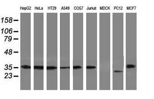
| Western blot analysis of extracts (35ug) from 9 different cell lines by using anti-ERCC1 monoclonal antibody (Clone 2E12). | |
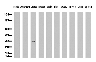
| Western Blot analysis of 10 different human tissue lysates (10ug) by using anti-ERCC1 monoclonal antibody (clone 2E12, 1:500) | |
| Immunohistochemical staining of paraffin-embedded Human breast tissue using anti-ERCC1 mouse monoclonal antibody. (Clone 2E12, dilution 1:100; heat-induced epitope retrieval by 10mM citric buffer, pH6.0, 120C for 3min) | |
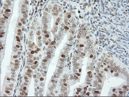
| Immunohistochemical staining of paraffin-embedded Human colon tissue using anti-ERCC1 mouse monoclonal antibody. (Clone 2E12, dilution 1:100; heat-induced epitope retrieval by 10mM citric buffer, pH6.0, 120C for 3min) | |
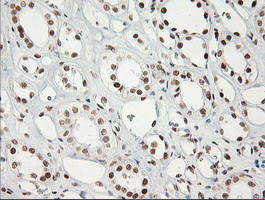
| Immunohistochemical staining of paraffin-embedded Human Kidney tissue using anti-ERCC1 mouse monoclonal antibody. (Clone 2E12, dilution 1:100; heat-induced epitope retrieval by 10mM citric buffer, pH6.0, 120C for 3min) | |
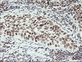
| Immunohistochemical staining of paraffin-embedded Carcinoma of Human kidney tissue using anti-ERCC1 mouse monoclonal antibody. (Clone 2E12, dilution 1:100; heat-induced epitope retrieval by 10mM citric buffer, pH6.0, 120C for 3min) | |
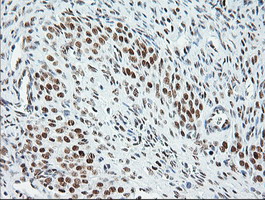
| Immunohistochemical staining of paraffin-embedded Carcinoma of Human lung tissue using anti-ERCC1 mouse monoclonal antibody. (Clone 2E12, dilution 1:100; heat-induced epitope retrieval by 10mM citric buffer, pH6.0, 120C for 3min) | |
| Immunohistochemical staining of paraffin-embedded Human Ovary tissue using anti-ERCC1 mouse monoclonal antibody. (Clone 2E12, dilution 1:100; heat-induced epitope retrieval by 10mM citric buffer, pH6.0, 120C for 3min) | |
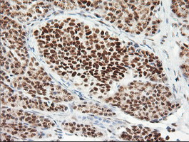
| Immunohistochemical staining of paraffin-embedded Adenocarcinoma of Human ovary tissue using anti-ERCC1 mouse monoclonal antibody. (Clone 2E12, dilution 1:100; heat-induced epitope retrieval by 10mM citric buffer, pH6.0, 120C for 3min) | |
| Immunohistochemical staining of paraffin-embedded Human pancreas tissue using anti-ERCC1 mouse monoclonal antibody. (Clone 2E12, dilution 1:100; heat-induced epitope retrieval by 10mM citric buffer, pH6.0, 120C for 3min) | |
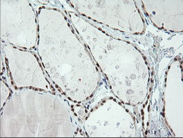
| Immunohistochemical staining of paraffin-embedded Human thyroid tissue using anti-ERCC1 mouse monoclonal antibody. (Clone 2E12, dilution 1:100; heat-induced epitope retrieval by 10mM citric buffer, pH6.0, 120C for 3min) | |
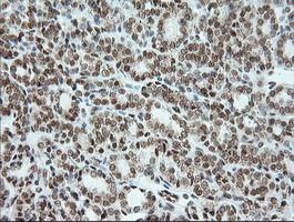
| Immunohistochemical staining of paraffin-embedded Carcinoma of Human thyroid tissue using anti-ERCC1 mouse monoclonal antibody. (Clone 2E12, dilution 1:100; heat-induced epitope retrieval by 10mM citric buffer, pH6.0, 120C for 3min) | |
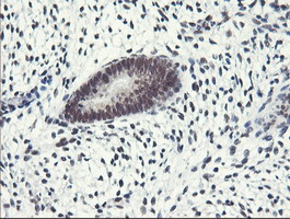
| Immunohistochemical staining of paraffin-embedded Human endometrium tissue using anti-ERCC1 mouse monoclonal antibody. (Clone 2E12, dilution 1:100; heat-induced epitope retrieval by 10mM citric buffer, pH6.0, 120C for 3min) | |
| Immunohistochemical staining of paraffin-embedded Adenocarcinoma of Human endometrium tissue using anti-ERCC1 mouse monoclonal antibody. (Clone 2E12, dilution 1:100; heat-induced epitope retrieval by 10mM citric buffer, pH6.0, 120C for 3min) | |
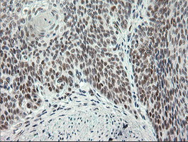
| Immunohistochemical staining of paraffin-embedded Human bladder tissue using anti-ERCC1 mouse monoclonal antibody. (Clone 2E12, dilution 1:100; heat-induced epitope retrieval by 10mM citric buffer, pH6.0, 120C for 3min) | |
| Immunohistochemical staining of paraffin-embedded Carcinoma of Human bladder tissue using anti-ERCC1 mouse monoclonal antibody. (Clone 2E12, dilution 1:100; heat-induced epitope retrieval by 10mM citric buffer, pH6.0, 120C for 3min) | |
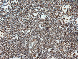
| Immunohistochemical staining of paraffin-embedded Human lymphoma tissue using anti-ERCC1 mouse monoclonal antibody. (Clone 2E12, dilution 1:100; heat-induced epitope retrieval by 10mM citric buffer, pH6.0, 120C for 3min) | |
| Immunohistochemical staining of paraffin-embedded Human tonsil using anti-ERCC1 mouse monoclonal antibody. (Clone 2E12, dilution 1:100; heat-induced epitope retrieval by 10mM citric buffer, pH6.0, 120C for 3min) | |
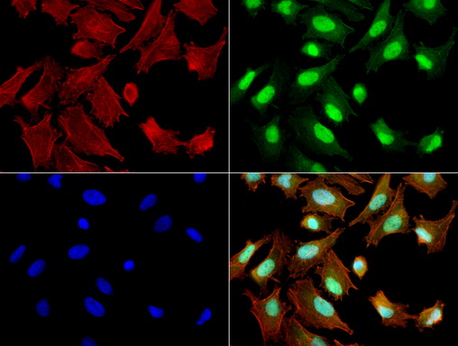
| Immunofluorescent staining of HeLa cells using ERCC1 mouse monoclonal antibody (UM500011, green). Actin filaments were labeled with TRITC-phalloidin (red), and nuclear with DAPI (blue). The three-color overlay image is located at the bottom-right corner. | |
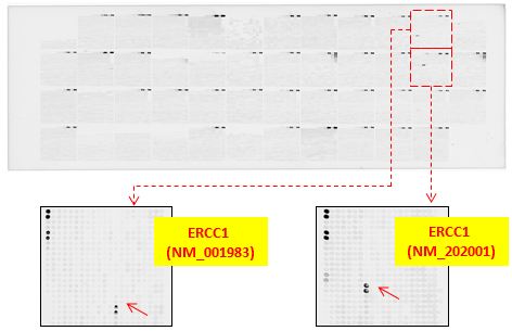
| OriGene overexpression protein microarray chip was immunostained with UltraMAB anti-ERCC1 mouse monoclonal antibody (Clone 2E12). The positive reactive proteins are highlighted with red arrows in the enlarged subarray. Other positive controls spotted in this subarray are serial dilutions of mouse IgG as controls. |


追加しました。
製品情報は掲載時点のものですが、価格表内の価格については随時最新のものに更新されます。お問い合わせいただくタイミングにより製品情報・価格などは変更されている場合があります。
表示価格に、消費税等は含まれていません。一部価格が予告なく変更される場合がありますので、あらかじめご了承下さい。