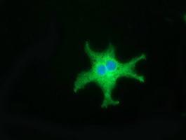
GTX84701 ICC/IF Image
Anti-CD80 mouse monoclonal antibody (GTX84701) immunofluorescent staining of COS7 cells transiently transfected with CD80

GTX84701 IHC-P Image
Immunohistochemical staining of paraffin-embedded Adenocarcinoma of Human breast tissue using anti-CD80 mouse monoclonal antibody. (GTX84701)
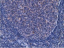
GTX84701 IHC-P Image
Immunohistochemical staining of paraffin-embedded Human lymph node tissue using anti-CD80 mouse monoclonal antibody. (GTX84701)
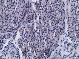
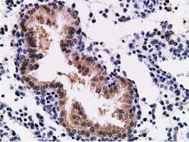
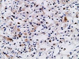
GTX84701 IHC-P Image
Immunohistochemical staining of paraffin-embedded Human pancreas tissue using anti-CD80 mouse monoclonal antibody. (GTX84701)
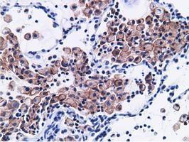
GTX84701 IHC-P Image
Immunohistochemical staining of paraffin-embedded Carcinoma of Human lung tissue using anti-CD80 mouse monoclonal antibody. (GTX84701)
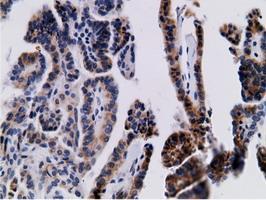
GTX84701 IHC-P Image
Immunohistochemical staining of paraffin-embedded Carcinoma of Human prostate tissue using anti-CD80 mouse monoclonal antibody. (GTX84701)
GTX84701 IHC-P Image
Immunohistochemical staining of paraffin-embedded Carcinoma of Human thyroid tissue using anti-CD80 mouse monoclonal antibody. (GTX84701)
GTX84701 IHC-P Image
Immunohistochemical staining of paraffin-embedded Adenocarcinoma of Human endometrium tissue using anti-CD80 mouse monoclonal antibody. (GTX84701)
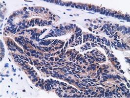
GTX84701 IHC-P Image
Immunohistochemical staining of paraffin-embedded Adenocarcinoma of Human ovary tissue using anti-CD80 mouse monoclonal antibody. (GTX84701)
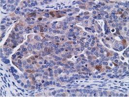
GTX84701 IHC-P Image
Immunohistochemical staining of paraffin-embedded Carcinoma of Human kidney tissue using anti-CD80 mouse monoclonal antibody. (GTX84701)
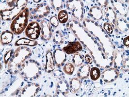
GTX84701 IHC-P Image
Immunohistochemical staining of paraffin-embedded Human Kidney tissue using anti-CD80 mouse monoclonal antibody. (GTX84701)
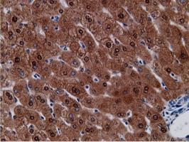
GTX84701 IHC-P Image
Immunohistochemical staining of paraffin-embedded Human liver tissue using anti-CD80 mouse monoclonal antibody. (GTX84701)
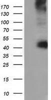
GTX84701 WB Image
HEK293T cells were transfected with the pCMV6-ENTRY control (Left lane) or pCMV6-ENTRY CD80 (Right lane) cDNA for 48 hrs and lysed. Equivalent amounts of cell lysates (5 ug per lane) were separated by SDS-PAGE and immunoblotted with anti-CD80.