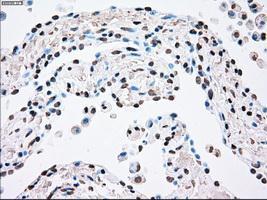
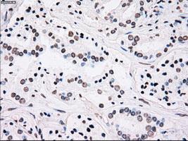
GTX83865 IHC-P Image
Immunohistochemical staining of paraffin-embedded endometrium tissue using anti-PLK1mouse monoclonal antibody. (GTX83865, Dilution 1:50)
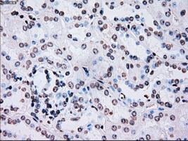
GTX83865 IHC-P Image
Immunohistochemical staining of paraffin-embedded Kidney tissue using anti-PLK1mouse monoclonal antibody. (GTX83865, Dilution 1:50)
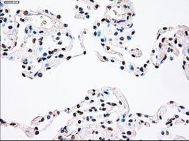
GTX83865 IHC-P Image
Immunohistochemical staining of paraffin-embedded lung tissue using anti-PLK1mouse monoclonal antibody. (GTX83865, Dilution 1:50)
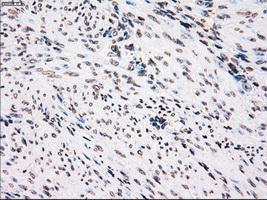
GTX83865 IHC-P Image
Immunohistochemical staining of paraffin-embedded Adenocarcinoma of breast tissue using anti-PLK1 mouse monoclonal antibody. (GTX83865, Dilution 1:50)
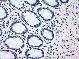
GTX83865 IHC-P Image
Immunohistochemical staining of paraffin-embedded colon tissue using anti-PLK1mouse monoclonal antibody. (GTX83865, Dilution 1:50)
GTX83865 IHC-P Image
Immunohistochemical staining of paraffin-embedded pancreas tissue using anti-PLK1mouse monoclonal antibody. (GTX83865, Dilution 1:50)
GTX83865 IHC-P Image
Immunohistochemical staining of paraffin-embedded Carcinoma of bladder tissue using anti-PLK1mouse monoclonal antibody. (GTX83865, Dilution 1:50)
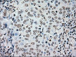
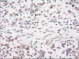
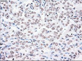
GTX83865 IHC-P Image
Immunohistochemical staining of paraffin-embedded Carcinoma of kidney tissue using anti-PLK1mouse monoclonal antibody. (GTX83865, Dilution 1:50)
GTX83865 IHC-P Image
Immunohistochemical staining of paraffin-embedded Carcinoma of lung tissue using anti-PLK1mouse monoclonal antibody. (GTX83865, Dilution 1:50)

GTX83865 IHC-P Image
Immunohistochemical staining of paraffin-embedded prostate tissue using anti-PLK1mouse monoclonal antibody. (GTX83865, Dilution 1:50)
GTX83865 IHC-P Image
Immunohistochemical staining of paraffin-embedded Adenocarcinoma of colon tissue using anti-PLK1mouse monoclonal antibody. (GTX83865, Dilution 1:50)
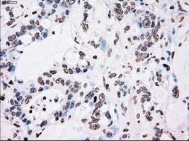
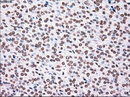
GTX83865 IHC-P Image
Immunohistochemical staining of paraffin-embedded Adenocarcinoma of endometrium tissue using anti-PLK1mouse monoclonal antibody. (GTX83865, Dilution 1:50)
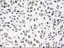
GTX83865 IHC-P Image
Immunohistochemical staining of paraffin-embedded Carcinoma of pancreas tissue using anti-PLK1mouse monoclonal antibody. (GTX83865, Dilution 1:50)
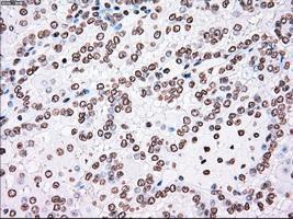
GTX83865 IHC-P Image
Immunohistochemical staining of paraffin-embedded Carcinoma of prostate tissue using anti-PLK1mouse monoclonal antibody. (GTX83865, Dilution 1:50)
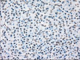
GTX83865 IHC-P Image
Immunohistochemical staining of paraffin-embedded Carcinoma of thyroid tissue using anti-PLK1mouse monoclonal antibody. (GTX83865, Dilution 1:50)
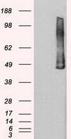
GTX83865 WB Image
HEK293T cells were transfected with the pCMV6-ENTRY control (Left lane) or pCMV6-ENTRY PLK1 (Right lane) cDNA for 48 hrs and lysed. Equivalent amounts of cell lysates (5 ug per lane) were separated by SDS-PAGE and immunoblotted with anti-PLK1.
 of PLK1 by using TrueMab monoclonal anti-PLK1 antibodies (Negative control: IP without adding anti-PLK1 antibody.). For each experiment, 500ul of DDK tagged PLK1 overexpression lysates (at 1:5 dilution with HEK293T lysate), 2ug of anti-PLK1 antibody and 20ul (0.1mg) of goat anti-mouse conjugated magnetic beads were mixed and incubated overnight. After extensive wash to remove any non-specific binding, the immuno-precipitated products were analyzed with rabbit anti-DDK polyclonal antibody.
)