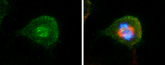
GTX70268 ICC/IF Image
Hec1 antibody [9G3.23] detects Hec1 protein at kinetochore by immunofluorescent analysis.Sample: HeLa cells were fixed in 4% paraformaldehyde at RT for 15 min.Green: Hec1 stained by Hec1 antibody [9G3.23] (GTX70268) diluted at 1:500.Red: alpha Tubulin 4a, a cytoskeleton marker, stained by alpha Tubulin 4a antibody (GTX112141) diluted at 1:500.Blue: Hoechst 33342 staining.
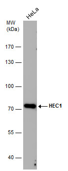
GTX70268 WB Image
Whole cell extract (30 ug) was separated by 7.5% SDS-PAGE, and the membrane was blotted with HEC1 antibody [9G3.23] (GTX70268) diluted at 1:1000. The signal was developed with Trident ECL plus-Enhanced.
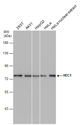
GTX70268 WB Image
HEC1 antibody [9G3.23] detects HEC1 protein by western blot analysis. Various whole cell extracts and HeLa nuclear extracts (30 ug) were separated by 7.5% SDS-PAGE, and the membrane was blotted with HEC1 antibody [9G3.23] (GTX70268) diluted at 1:1000. The HRP-conjugated anti-mouse IgG antibody (GTX213111-01) was used to detect the primary antibody, and the signal was developed with Trident femto Western HRP Substrate (GTX14698).

GTX70268 ICC/IF Image
Mouse anti-Hec1 (9G3, cat# GTX 70268) and rabbit anti-Hec1 (phospho Ser165) antibodies were used to co-stain MCF10A cells. Insets show kinetochores of misaligned chromosomes. DAPI staining shows chromatin.
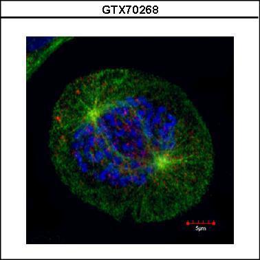
GTX70268 ICC/IF Image
Confocal immunofluorescence staining (Olympus FV10i) of HEC1, a kinetochore outer layer marker. U2OS cells were fixed by 4% PFA and costained with Hec1 9G3.23 AB (Red; cat# GTX70268; 1:500 Ab dilution) and alpha-tubulin rabbit polyclonal AB (Green; cat# GTX112141; 1:500 Ab dilution ), a spindle marker. DAPI (blue), chromosomes. Scale bar, 5 um