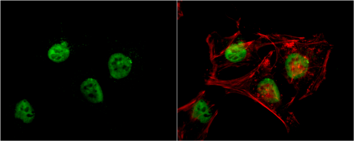
GTX70234 ICC/IF Image
RbAp48 antibody [13D10] detects RbAp48 protein at nucleus by immunofluorescent analysis.
Sample: HeLa cells were fixed in 4% paraformaldehyde at RT for 15 min.
Green: RbAp48 protein stained by RbAp48 antibody [13D10] (GTX70234) diluted at 1:200.
Red: phalloidin, a cytoskeleton marker, stained by phalloidin (invitrogen, A12380) diluted at 1:200.
Blue: Hoechst 33342 staining.
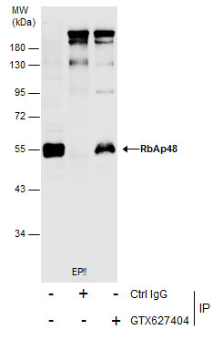
GTX70234 IP Image
Immunoprecipitation of RbAp48 protein from HeLa whole cell extract using 5 ug of RbAp48 antibody [13D10] (GTX70234).
Western blot analysis was performed using RbAp48 antibody [13D10] (GTX70234).
EasyBlot HRP-conjugated anti mouse IgG antibody (GTX221667-01) was used to detect the primary antibody, and the signal was developed with Trident ECL plus-Enhanced.
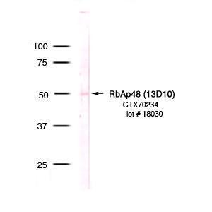
GTX70234 WB Image
Detection of RbAp48 in HeLa nuclear extract (GTX24105)
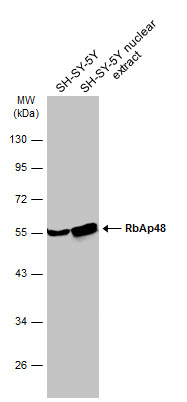
GTX70234 WB Image
SH-SY-5Y whole cell and nuclear extracts (30 ug) were separated by 10% SDS-PAGE, and the membrane was blotted with RbAp48 antibody [13D10] (GTX70234) diluted at 1:2000. The HRP-conjugated anti-mouse IgG antibody (GTX213111-01) was used to detect the primary antibody.