
GTX70020 IHC-P Image
Immunohistochemical analysis of paraffin-embedded cervical CA tissue sections using anti-CAIX antibody [GT12] (GTX70020) at a dilution of 1:1000. The hypoxic regions of the tumor show positive CAIX staining.

GTX70020 IHC-P Image
Immunohistochemical analysis of paraffin-embedded renal cell carcinoma (clear cell type) using anti-CAIX antibody [GT12] (GTX70020) at a dilution of 1:1000.
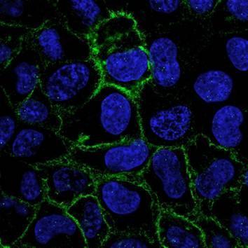
GTX70020 ICC/IF Image
Confocal immunofluorescence analysis (Olympus FV10i) of methanol-fixed A431 cells treated with 200uM CoCl2 for 48hr using anti-CAIX antibody [GT12] (GTX70020) at a dilution of 1:1000.

GTX70020 IHC-P Image
Immunohistochemical analysis of paraffin-embedded cervical CA tissue sections using anti-CAIX antibody [GT12] (GTX70020) at a dilution of 1:1000. The hypoxic regions of the tumor show positive CAIX staining.
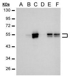
GTX70020 WB Image
Sample (30 ug HeLa whole cell lysate)
A: 24 hr Untreated
B: 24 hr treatment with 100uM CoCl2
C: 24 hr treatment with 200uM CoCl2
D: 48 hr Untreated
E: 48 hr treatment with 100uM CoCl2
F: 48 hr treatment with 200uM CoCl2
Anti-CAIX antibody [GT12] (GTX70020) was used at a dilution of 1:1000
The HRP-conjugated anti-mouse IgG antibody (GTX213111-01) was used to detect the primary antibody.
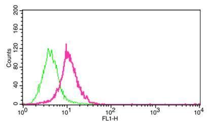
GTX70020 FACS Image
Flow cytometry on HeLa cells (1x10^6) stained with anti-CAIX antibody [GT12] (GTX70020) at a 1:1000 dilution. HeLa cells were untreated (green) or treated with 200uM CoCl2 (pink) for 48 hr.