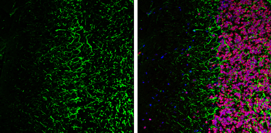
GTX634289 IHC-Fr Image
NF-H antibody [GT114] detects NF-H protein expression by immunohistochemical analysis.
Sample: Frozen-sectioned adult mouse cerebellum.
Green: NF-H protein stained by NF-H antibody [GT114] (GTX634289) diluted at 1:250.
Red: NeuN, stained by NeuN antibody (GTX133127) diluted at 1:500.
Blue: Fluoroshield with DAPI (GTX30920).
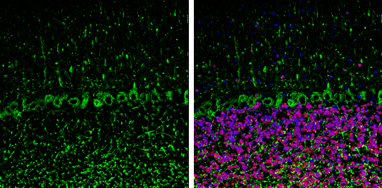
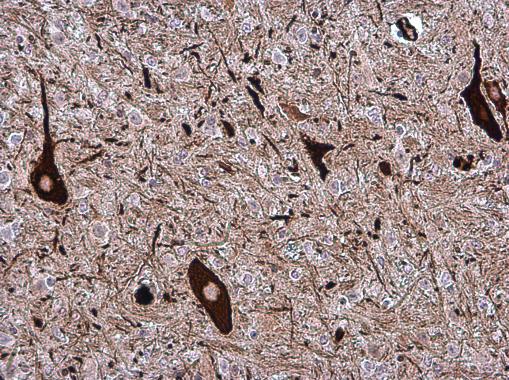
GTX634289 IHC-P Image
NF-H antibody [GT114] detects NF-H protein at cytoplasm in mouse brain by immunohistochemical analysis.
Sample: Paraffin-embedded mouse brain.
NF-H antibody [GT114] (GTX634289) diluted at 1:500.
Scale bar = 1 um.
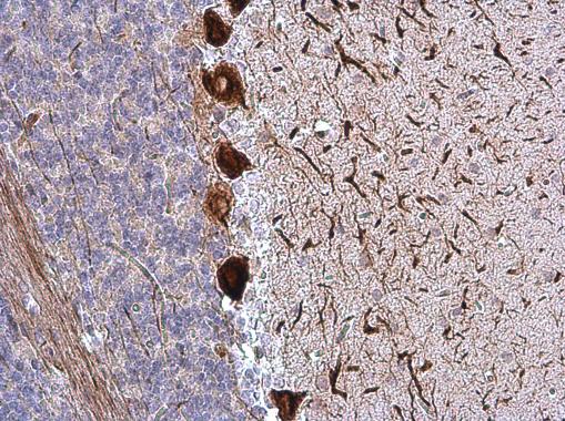
GTX634289 IHC-P Image
NF-H antibody [GT114] detects NF-H protein at cytoplasm in rat brain by immunohistochemical analysis.
Sample: Paraffin-embedded rat brain.
NF-H antibody [GT114] (GTX634289) diluted at 1:500.
Scale bar = 1 um.
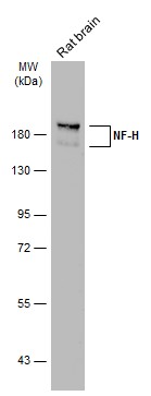
GTX634289 WB Image
Rat tissue extract (50 ug) was separated by 7.5% SDS-PAGE, and the membrane was blotted with NF-H antibody (GTX634289) diluted at 1:1000. The HRP-conjugated anti-mouse IgG antibody (GTX213111-01) was used to detect the primary antibody.
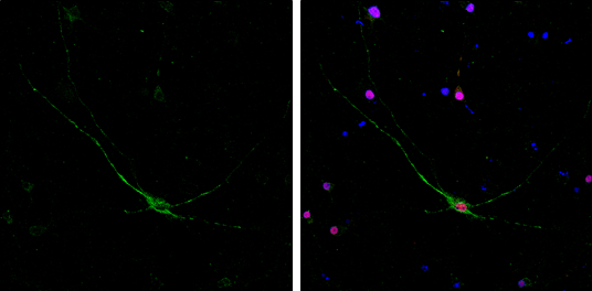
GTX634289 ICC/IF Image
NF-H antibody [GT114] detects NF-H protein by immunofluorescent analysis.
Sample: DIV10 rat E18 primary cortical neurons were fixed in 4% paraformaldehyde at RT for 15 min.
Green: NF-H protein stained by NF-H antibody [GT114] (GTX634289) diluted at 1:500.
Red: NeuN, stained by NeuN antibody (GTX132974) diluted at 1:250.
Blue: Fluoroshield with DAPI (GTX30920).
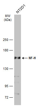
GTX634289 WB Image
Whole cell extract (30 ug) was separated by 5% SDS-PAGE, and the membrane was blotted with NF-H antibody [GT114] (GTX634289) diluted at 1:1000. The HRP-conjugated anti-mouse IgG antibody (GTX213111-01) was used to detect the primary antibody.
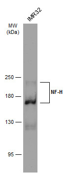
GTX634289 WB Image
Whole cell extract (30 ug) was separated by 5% SDS-PAGE, and the membrane was blotted with NF-H antibody [GT114] (GTX634289) diluted at 1:1000. The HRP-conjugated anti-mouse IgG antibody (GTX213111-01) was used to detect the primary antibody.