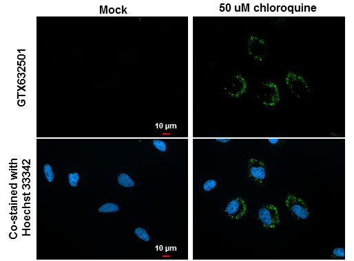
GTX632501 ICC/IF Image
LC3B antibody [GT3612] detects LC3B protein at autophagosome by immunofluorescent analysis.
Samples: HeLa cells mock (left) and treated with 50uM Chloroquine for 24 hr (right) were fixed in 4% paraformaldehyde at RT for 15 min.
Green: LC3B protein stained by LC3B antibody [GT3612] (GTX632501) diluted at 1:200.
Blue: Hoechst 33342 staining.
Scale bar = 10 um.
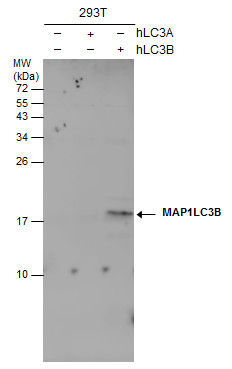
GTX632501 WB Image
Non-transfected (?) and transfected (+) 293T whole cell extracts (30 ug) were separated by 15% SDS-PAGE, and the membrane was blotted with LC3B antibody [GT3612] (GTX632501) diluted at 1:500.
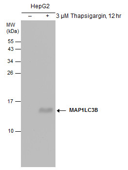
GTX632501 WB Image
Untreated (?) and treated (+) HepG2 whole cell extracts (30 ug) were separated by 15% SDS-PAGE, and the membrane was blotted with LC3B antibody [GT3612] (GTX632501) diluted at 1:500.
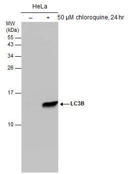
GTX632501 WB Image
Untreated (?) and treated (+) HeLa whole cell extracts (50 ug) were separated by 15% SDS-PAGE, and the membrane was blotted with LC3B antibody [GT3612] (GTX632501) diluted at 1:500.