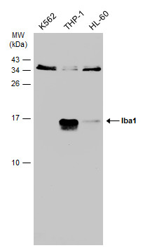
GTX632426 WB Image
Various whole cell extracts (30 ug) were separated by 15% SDS-PAGE, and the membrane was blotted with Iba1 antibody [GT10312] (GTX632426) diluted at 1:500. The HRP-conjugated anti-mouse IgG antibody (GTX213111-01) was used to detect the primary antibody.
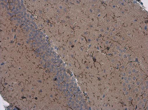
GTX632426 IHC-P Image
Iba1 antibody [GT10312] detects Iba1 protein at microglia in mouse brain by immunohistochemical analysis.
Sample: Paraffin-embedded mouse brain.
Iba1 antibody [GT10312] (GTX632426) diluted at 1:200.
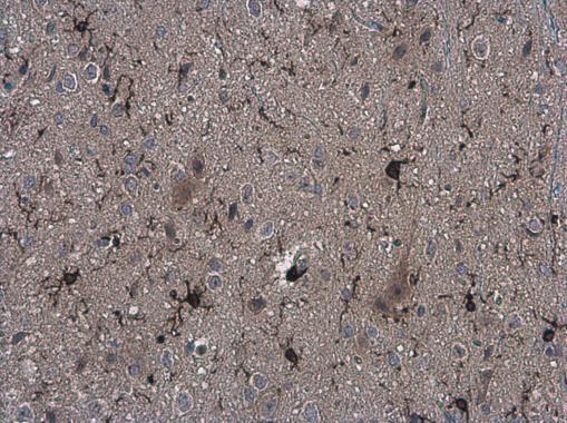
GTX632426 IHC-P Image
Iba1 antibody [GT10312] detects Iba1 protein at microglia in rat brain by immunohistochemical analysis.
Sample: Paraffin-embedded rat brain.
Iba1 antibody [GT10312] (GTX632426) diluted at 1:200.
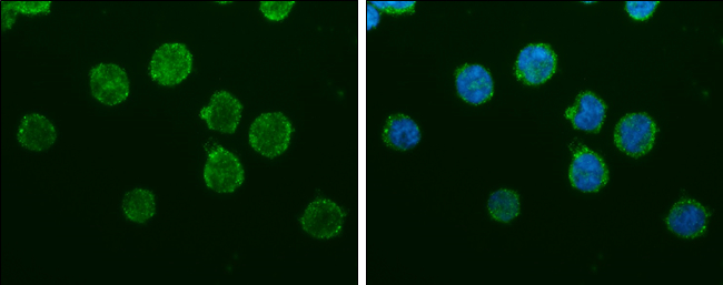
GTX632426 ICC/IF Image
Iba1 antibody [GT10312] detects Iba1 protein at cytoplasm by immunofluorescent analysis.
Sample: THP-1 cells were fixed in 4% paraformaldehyde at RT for 15 min.
Green: Iba1 protein stained by Iba1 antibody [GT10312] (GTX632426) diluted at 1:200.
Blue: Hoechst 33342 staining.
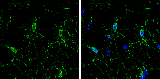
GTX632426 IHC-Fr Image
Iba1 antibody [GT10312] detects Iba1 protein by immunohistochemical analysis.
Samples: Frozen Sectioned adult mouse hippocampus.
Green: Iba1 protein stained by Iba1 antibody [GT10312] (GTX632426) diluted at 1:250.
Blue: Fluoroshield with DAPI (GTX30920).