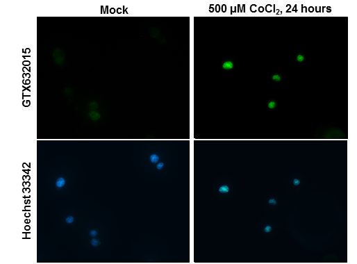
GTX632015 ICC/IF Image
HIF2 alpha antibody [GT125] detects HIF2 alpha protein at nucleus by immunofluorescent analysis.
Samples: PC-12 cells mock (left) and treated with 500 uM CoCl2 for 24 hrs (right) were fixed in 4% paraformaldehyde at RT for 15 min.
Green: HIF2 alpha protein stained by HIF2 alpha antibody [GT125] (GTX632015) diluted at 1:100.
Blue: Hoechst 33342 staining.

GTX632015 IP Image
Immunoprecipitation of HIF2 alpha protein from HepG2 whole cell extracts treated with 500 uM CoCl2 for 24 hr using 5 ug of HIF2 alpha antibody [GT125] (GTX632015).
Western blot analysis was performed using HIF2 alpha antibody [GT125] (GTX632015).
EasyBlot anti-Mouse IgG (GTX221667-01) was used as a secondary reagent.
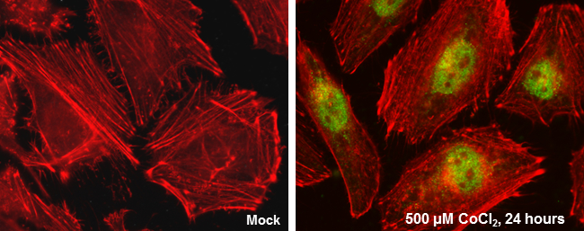
GTX632015 ICC/IF Image
HIF2 alpha antibody [GT125] detects HIF2 alpha protein at nucleus by immunofluorescent analysis.
Sample: HeLa cells were fixed in 4% paraformaldehyde at RT for 15 min.
Green: HIF2 alpha protein stained by HIF2 alpha antibody [GT125] (GTX632015) diluted at 1:100.
Red: Actin filaments were labeled with Phalloidin.
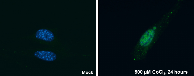
GTX632015 ICC/IF Image
HIF2 alpha antibody [GT125] detects HIF2 alpha protein at nucleus by immunofluorescent analysis.
Sample: NIH/3T3 cells were fixed in 4% paraformaldehyde at RT for 15 min.
Green: HIF2 alpha protein stained by HIF2 alpha antibody [GT125] (GTX632015) diluted at 1:100.
Blue: Hoechst 33342 staining.
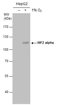
GTX632015 WB Image
HIF2 alpha antibody detects HIF2 alpha protein by western blot analysis. Un-treated (-) and treated (+, 1% O2 treatment for 24hr) HepG2 whole cell extracts (30 ug) were separated by 7.5% SDS-PAGE, and the membrane was blotted with HIF2 alpha antibody (GTX632015) at a dilution of 1:500. The HRP-conjugated anti-mouse IgG antibody (GTX213111-01) was used to detect the primary antibody.