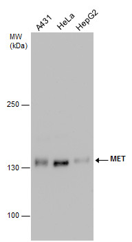
GTX631992 WB Image
c-Met antibody detects c-Met protein by western blot analysis. Various whole cell extracts (30 ug) were separated by 5% SDS-PAGE, and the membrane was blotted with c-Met antibody (GTX631992) diluted by 1:1000.
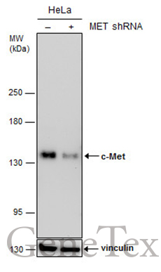
GTX631992 WB Image
Non-transfected (?) and transfected (+) HeLa whole cell extracts (30 ug) were separated by 5% SDS-PAGE, and the membrane was blotted with c-Met antibody [GT556] (GTX631992) diluted at 1:500.
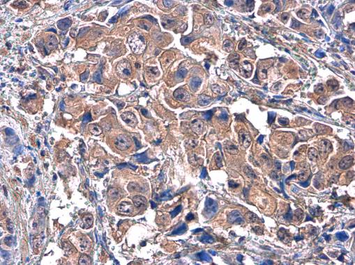
GTX631992 IHC-P Image
c-Met antibody [GT556] detects c-Met protein at cytoplasm in human breast carcinoma by immunohistochemical analysis.
Sample: Paraffin-embedded human breast carcinoma.
c-Met antibody [GT556] (GTX631992) diluted at 1:100.
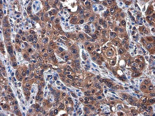
GTX631992 IHC-P Image
c-Met antibody [GT556] detects c-Met protein at cytoplasm in human esophageal carcinoma by immunohistochemical analysis.
Sample: Paraffin-embedded human esophageal carcinoma.
c-Met antibody [GT556] (GTX631992) diluted at 1:100.
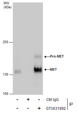
GTX631992 IP Image
Immunoprecipitation of c-Met protein from HeLa whole cell extracts using 5 ug of MET antibody [GT556] (GTX631992).
Western blot analysis was performed using c-Met antibody [GT556] (GTX631992).
EasyBlot anti-Mouse IgG (GTX221667-01) was used as a secondary reagent.
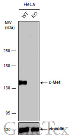
GTX631992 WB Image
Wild-type (WT) and c-Met knockout (KO) HeLa cell extracts (30 ug) were separated by 5% SDS-PAGE, and the membrane was blotted with c-Met antibody [GT556] (GTX631992) diluted at 1:1000. The HRP-conjugated anti-mouse IgG antibody (GTX213111-01) was used to detect the primary antibody, and the signal was developed with Trident ECL plus-Enhanced.