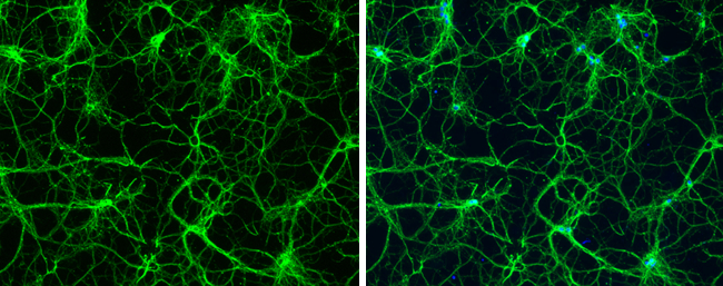
GTX631836 ICC/IF Image
beta Tubulin 3/ TUJ1 antibody [GT11710] detects beta Tubulin 3/ TUJ1 protein expression by immunofluorescent analysis.
Sample: Cultured rat E18 primary hippocampal neuron. Cells were fixed in 4% paraformaldehyde at RT for 15 min.
Green: beta Tubulin 3/ TUJ1 protein stained by beta Tubulin 3/ TUJ1 antibody [GT11710] (GTX631836) diluted at 1:250.
Blue: Fluoroshield with DAPI (GTX30920).
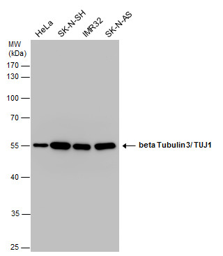
GTX631836 WB Image
beta Tubulin 3/ TUJ1 antibody detects beta Tubulin 3/ TUJ1 protein by western blot analysis. Various whole cell extracts (20 ug) were separated by 10% SDS-PAGE, and the membrane was blotted with beta Tubulin 3/ TUJ1 antibody (GTX631836) diluted at a dilution of 1:1000. The HRP-conjugated anti-mouse IgG antibody (GTX213111-01) was used to detect the primary antibody.
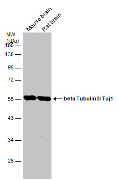
GTX631836 WB Image
Various tissue extracts (10 ug) were separated by 10% SDS-PAGE, and the membrane was blotted with beta Tubulin 3/ Tuj1 antibody [GT11710] (GTX631836) diluted at 1:20000. The HRP-conjugated anti-mouse IgG antibody (GTX213111-01) was used to detect the primary antibody.

GTX631836 IHC-P Image
beta Tubulin 3/ TUJ1 antibody [GT11710] detects beta Tubulin 3/ TUJ1 protein at cytoplasm in rat brain by immunohistochemical analysis.
Sample: Paraffin-embedded rat brain.
beta Tubulin 3/ TUJ1 antibody [GT11710] (GTX631836) diluted at 1:500.
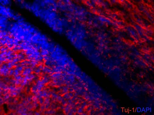
GTX631836 IHC-Fr Image
beta III Tubulin antibody [GT11710] detects beta III Tubulin proteins on embryonic mouse brain by immunohistochemical analysis.
Sample:Frozen section of embryonic mouse brain (mE18.5).
Red: beta III Tubulin antibody [GT11710] (GTX631836) diluted at 1:500.
Blue: DAPI
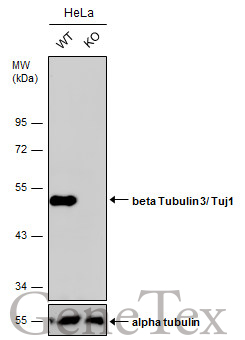
GTX631836 WB Image
Wild-type (WT) and beta Tubulin 3/ Tuj1 knockout (KO) HeLa cell extracts (30 ug) were separated by 10% SDS-PAGE, and the membrane was blotted with beta Tubulin 3/ Tuj1 antibody [GT11710] (GTX631836) diluted at 1:2500. The HRP-conjugated anti-mouse IgG antibody (GTX213111-01) was used to detect the primary antibody, and the signal was developed with Trident ECL plus-Enhanced.
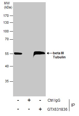
GTX631836 IP Image
Immunoprecipitation of beta III Tubulin protein from SK-N-SH whole cell extracts using 5 ug of beta III Tubulin antibody [GT11710] (GTX631836).
Western blot analysis was performed using beta III Tubulin antibody [GT11710] (GTX631836).
EasyBlot anti-Mouse IgG (GTX221667-01) was used as a secondary reagent.
GTX631836 IHC-Fr Image
beta Tubulin 3/ TUJ1 antibody [GT11710] detects beta Tubulin 3/ TUJ1 protein by immunohistochemical analysis.
Sample: Frozen sectioned E13.5 rat brain.
Green: SOX2 protein stained by SOX2 antibody [N1C3] (GTX101507) diluted at 1:250.
Red: beta Tubulin 3/ TUJ1 protein stained by beta Tubulin 3/ TUJ1 antibody [GT11710] (GTX131836) diluted at 1:250.
Blue: Fluoroshield with DAPI (GTX30920).
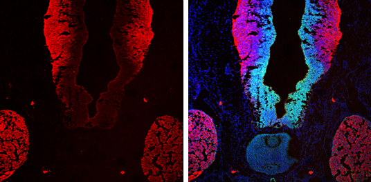
GTX631836 IHC-Fr Image
beta Tubulin 3/ TUJ1 antibody [GT11710] detects beta Tubulin 3/ TUJ1 protein by immunohistochemical analysis.
Sample: Frozen sectioned E13.5 rat brain.
Red: beta Tubulin 3/ TUJ1 protein stained by beta Tubulin 3/ TUJ1 antibody [GT11710] (GTX131836) diluted at 1:250.
Blue: Fluoroshield with DAPI (GTX30920).
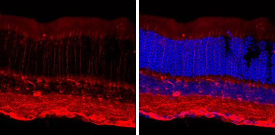
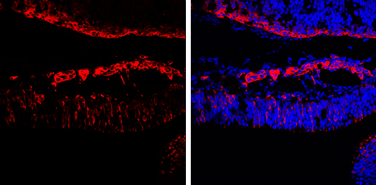
GTX631836 IHC-Fr Image
beta Tubulin 3/ TUJ1 antibody [GT11710] detects beta Tubulin 3/ TUJ1 protein by immunohistochemical analysis.
Sample: Frozen sectioned adult mouse retina.
Red: beta Tubulin 3/ TUJ1 protein stained by beta Tubulin 3/ TUJ1 antibody [GT11710] (GTX631836) diluted at 1:250.
Blue: Fluoroshield with DAPI (GTX30920).