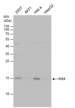
GTX630983 WB Image
FIS1 antibody detects FIS1 protein by western blot analysis. Various whole cell extracts (30 ug) were separated by 15% SDS-PAGE, and the membrane was blotted with FIS1 antibody (GTX630983) diluted by 1:1000.
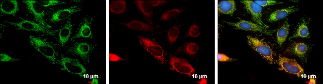
GTX630983 ICC/IF Image
FIS1 antibody detects FIS1 protein at mitochondria by immunofluorescent analysis.
Sample: HeLa cells were fixed in 2% paraformaldehyde/culture medium at 37oC for 30 min.
Green: FIS1 protein stained by FIS1 antibody (GTX630983) diluted at 1:1000.
Red: MitoTrackerR Red CMXRos, a mitochondria tracker.
Blue: Hoechst 33342 staining.
Scale bar = 10 um.
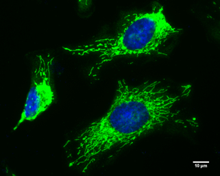
GTX630983 ICC/IF Image
FIS1 antibody [GT4211] detects FIS1 protein at mitochondria by immunofluorescent analysis.
Sample: HeLa cells were fixed in 4% paraformaldehyde at room temperature.
Green: FIS1 protein stained by FIS1 antibody [GT4211] (GTX630983) diluted at 1:100.
Blue: Hoechst 33342 staining.
Scale bar = 10 um.
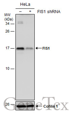
GTX630983 WB Image
Non-transfected (?) and transfected (+) HeLa whole cell extracts (30 ug) were separated by 15% SDS-PAGE, and the membrane was blotted with FIS1 antibody [GT4211] (GTX630983) diluted at 1:500.