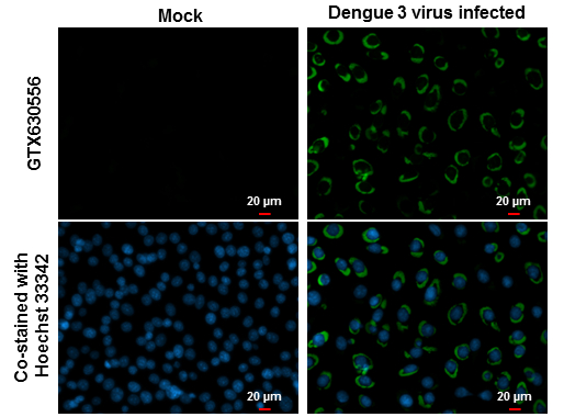
GTX630556 ICC/IF Image
NS1 (Dengue virus ) antibody [GT4212] detects NS1 (Dengue virus ) protein at cytoplasm by immunofluorescent analysis.
Samples: BHK-21 cells mock (left) and infected with Dengue virus 3 (right) were fixed in MeOH.
Green: NS1 (Dengue virus ) protein stained by NS1 (Dengue virus ) antibody [GT4212] (GTX630556) diluted at 1:1000.
Blue: Hoechst 33342 staining.
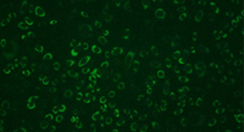
GTX630556 ICC/IF Image
NS1 (DEN) antibody [GT4212] (GTX630556) mouse mAb. IFA of MeOH/Acetone-fixed DEN2-infected BHK-21 cells (1:1000).
Green: non-structural protein 1 (Dengue virus 2) protein stained by NS1 (Dengue virus ) antibody [GT4212] (GTX630556) diluted at 1:1000.
Blue: DAPI staining.
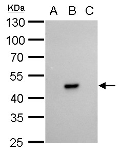
GTX630556 WB Image
NS1 (Dengue virus ) antibody [GT4212] detects NS1 (Dengue virus ) protein by western blot analysis.
A. 30 ug BHK21 whole cell lysate/extract
B. 30 ug whole cell lysate/extract of Dengue virus type 2 infected BHK21 cells
C. 30 ug whole cell lysate/extract of Japanese encephalitis virus infected BHK21 cells
10% SDS-PAGE
NS1 (Dengue virus ) antibody [GT4212] (GTX630556) dilution: 1:20000
The HRP-conjugated anti-mouse IgG antibody (GTX213111-01) was used to detect the primary antibody.
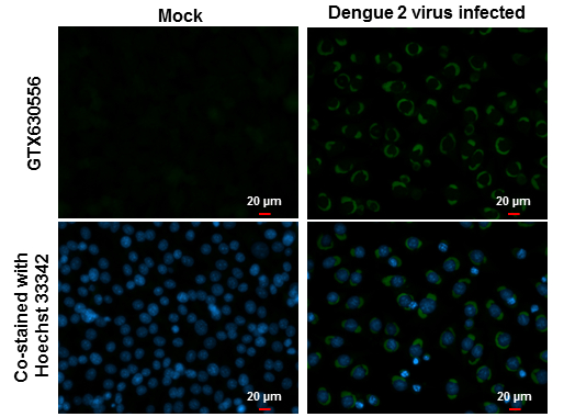
GTX630556 ICC/IF Image
NS1 (Dengue virus ) antibody [GT4212] detects NS1 (Dengue virus ) protein at cytoplasm by immunofluorescent analysis.
Samples: BHK-21 cells mock (left) and infected with Dengue virus 2 (right) were fixed in MeOH.
Green: NS1 (Dengue virus ) protein stained by NS1 (Dengue virus ) antibody [GT4212] (GTX630556) diluted at 1:1000.
Blue: Hoechst 33342 staining.