GTX630413 ICC/IF Image
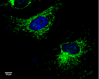
UQCRC1 antibody [GT139] detects UQCRC1 protein at mitochondria by immunofluorescent analysis.
Sample: HeLa cells were fixed in 4% paraformaldehyde at room temperature.
Green: UQCRC1 protein stained by UQCRC1 antibody [GT139] (GTX630413) diluted at 1:100.
Blue: Hoechst 33342 staining.
Scale bar = 10 um.
GTX630413 WB Image
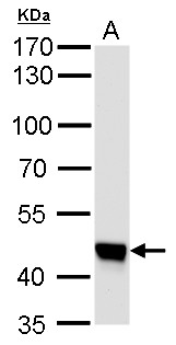
UQCRC1 antibody [GT139] detects UQCRC1 protein by western blot analysis.
A. 50 ug mouse brain lysate/extract
7.5 % SDS-PAGE
UQCRC1 antibody [GT139] (GTX630413) dilution: 1:1000
GTX630413 WB Image
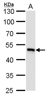
UQCRC1 antibody [GT139] detects UQCRC1 protein by western blot analysis.
A. 50 ug rat brain lysate/extract
10 % SDS-PAGE
UQCRC1 antibody [GT139] (GTX630413) dilution: 1:1000
GTX630413 WB Image
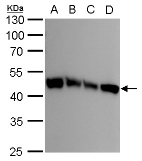
UQCRC1 antibody [GT139] detects UQCRC1 protein by western blot analysis.
A. 30 ug 293T whole cell lysate/extract
B. 30 ug A431 whole cell lysate/extract
C. 30 ug HeLa whole cell lysate/extract
D. 30 ug HepG2 whole cell lysate/extract
10 % SDS-PAGE
UQCRC1 antibody [GT139] (GTX630413) dilution: 1:1000
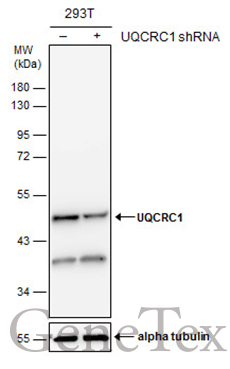
GTX630413 WB Image
Non-transfected (?) and transfected (+) 293T whole cell extracts (30 ug) were separated by 10% SDS-PAGE, and the membrane was blotted with UQCRC1 antibody [GT139] (GTX630413) diluted at 1:500.