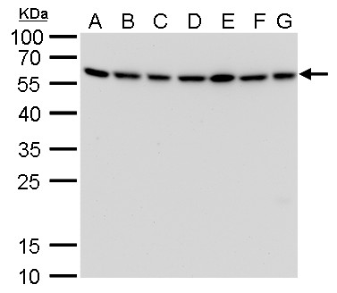
GTX629815 WB Image
ATG12 antibody [GT166] detects ATG12 protein by western blot analysis.
A. 30 ug Neuro2A whole cell lysate/extract
B. 30 ug GL261 whole cell lysate/extract
C. 30 ug C8D30 whole cell lysate/extract
D. 30 ug NIH-3T3 whole cell lysate/extract
E. 30 ug BCL-1 whole cell lysate/extract
F. 30 ug Raw264.7 whole cell lysate/extract
G. 30 ug C2C12 whole cell lysate/extract
12% SDS-PAGE
ATG12 antibody [GT166] (GTX629815) dilution: 1:1000
The HRP-conjugated anti-mouse IgG antibody (GTX213111-01) was used to detect the primary antibody.
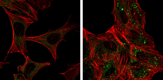
GTX629815 ICC/IF Image
ATG12 antibody [GT166] detects ATG12 protein at autophagosome by immunofluorescent analysis.
Samples: HeLa cells mock (left) and treated with 50uM Chloroquine for 24 hr (right) were fixed in 4% paraformaldehyde at RT for 15 min.
Green: ATG12 protein stained by ATG12 antibody [GT166] (GTX629815) diluted at 1:1000.
Red: Phalloidin, a F-actin marker.
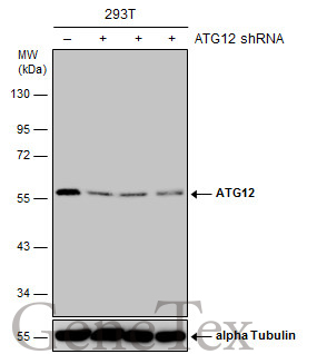
GTX629815 WB Image
Non-transfected (?) and transfected (+) 293T whole cell extracts (30 ug) were separated by 10% SDS-PAGE, and the membrane was blotted with ATG12 antibody [GT166] (GTX629815) diluted at 1:500. The HRP-conjugated anti-mouse IgG antibody (GTX213111-01) was used to detect the primary antibody.
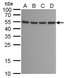
GTX629815 WB Image
ATG12 antibody [GT166] detects ATG12 protein by western blot analysis.
A. 30 ug 293T whole cell lysate/extract
B. 30 ug A431 whole cell lysate/extract
C. 30 ug HeLa whole cell lysate/extract
D. 30 ug HepG2 whole cell lysate/extract
12% SDS-PAGE
ATG12 antibody [GT166] (GTX629815) dilution: 1:1000
The HRP-conjugated anti-mouse IgG antibody (GTX213111-01) was used to detect the primary antibody.