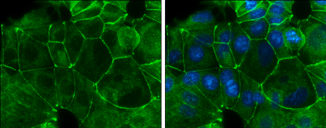
GTX629694 ICC/IF Image
E-Cadherin antibody [GT358] detects E-Cadherin protein at cell membrane by immunofluorescent analysis.
Sample: MCF7 cells were fixed in ice-cold MeOH for 5 min.
Green: E-Cadherin protein stained by E-Cadherin antibody [GT358] (GTX629694) diluted at 1:500.
Blue: Hoechst 33342 staining.
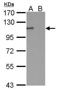
GTX629694 WB Image
E-cadherin antibody [GT358] detects E-cadherin protein by Western blot analysis.
A. 30 ug MCF-7 whole cell lysate/extract
B. 30 ug MDA-MB-231 whole cell lysate/extract
7.5 % SDS-PAGE
E-cadherin antibody [GT358] (GTX629694) dilution: 1:1000
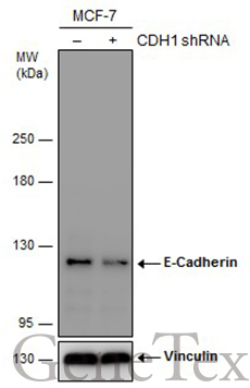
GTX629694 WB Image
Non-transfected (?) and transfected (+) MCF-7 whole cell extracts (30 ug) were separated by 5% SDS-PAGE, and the membrane was blotted with E-Cadherin antibody [GT358] (GTX629694) diluted at 1:500. The HRP-conjugated anti-mouset IgG antibody (GTX213111-01) was used to detect the primary antibody.