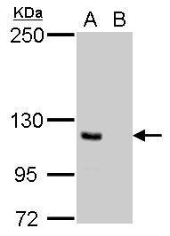
GTX629691 IP Image
E-adherin antibody [GT477] immunoprecipitates E-adherin protein in IP experiments.
IP samples: MCF-7 whole cell extract
A. Control with 3 ug of preimmune Mouse IgG
B. Immunoprecipitation of E-adherin protein by 3 ug E-adherin antibody [GT477] (GTX629691)
5 % SDS-PAGE
The immunoprecipitated E-adherin protein was detected by E-adherin antibody [GT477] (GTX629691) diluted at 1:500.
[EasyBlot anti-mouse IgG (GTX221667-01) was used as a secondary reagent]
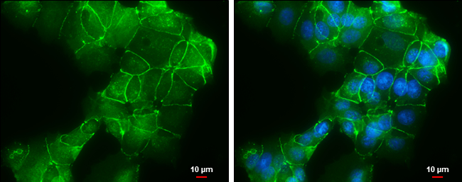
GTX629691 ICC/IF Image
E-Cadherin antibody [GT477] detects E-Cadherin protein at cell membrane by immunofluorescent analysis.
Sample: MCF7 cells were fixed in ice-cold MeOH for 5 min.
Green: E-Cadherin protein stained by E-Cadherin antibody [GT477] (GTX629691) diluted at 1:500.
Blue: Hoechst 33342 staining.
Scale bar = 10 um.
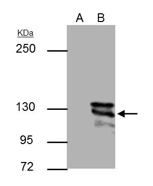
GTX629691 WB Image
E-cadherin antibody [GT477] detects E-cadherin protein by Western blot analysis.
A. 30 ug MCF-7 whole cell lysate/extract
B. 30 ug MDA-MB-231 whole cell lysate/extract
5 % SDS-PAGE
E-cadherin antibody [GT477] (GTX629691) dilution: 1:1000
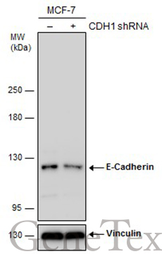
GTX629691 WB Image
Non-transfected (?) and transfected (+) MCF-7 whole cell extracts (30 ug) were separated by 5% SDS-PAGE, and the membrane was blotted with E-Cadherin antibody [GT477] (GTX629691) diluted at 1:2000. The HRP-conjugated anti-mouset IgG antibody (GTX213111-01) was used to detect the primary antibody.
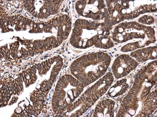
GTX629691 IHC-P Image
E-Cadherin antibody [GT477] detects E-Cadherin protein at cell membrane and cytoplasm in mouse intestine by immunohistochemical analysis.
Sample: Paraffin-embedded mouse intestine.
E-Cadherin antibody [GT477] (GTX629691) diluted at 1:250.
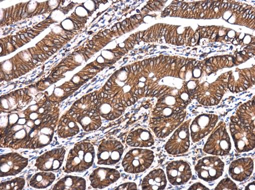
GTX629691 IHC-P Image
E-Cadherin antibody [GT477] detects E-Cadherin protein at cell membrane and cytoplasm in rat intestine by immunohistochemical analysis.
Sample: Paraffin-embedded rat intestine.
E-Cadherin antibody [GT477] (GTX629691) diluted at 1:250.