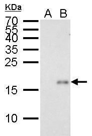
GTX629627 WB Image
p21 Cip1 antibody [GT8611] detects p21 Cip1 protein by Western blot analysis.
A. 30 ug HCT116 whole cell lysate/extract (untreated)
B. 30 ug HCT116 whole cell lysate/extract (30 uM Cisplatin treatment for 24 hr)
15 % SDS-PAGE
p21 Cip1 antibody [GT8611] (GTX629627) dilution: 1:1000
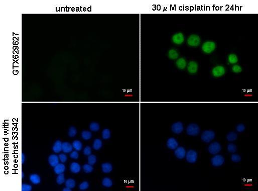
GTX629627 ICC/IF Image
p21 Cip1 antibody [GT8611] detects p21 Cip1 protein at nucleus by immunofluorescent analysis.
Sample: HCT116 cells treated with 30uM cisplatin for 24hr and were fixed in 4% paraformaldehyde at RT for 15 min.
Green: p21 Cip1 protein stained by p21 Cip1 antibody [GT8611] (GTX629627) diluted at 1:500.
Blue: Hoechst 33342 staining.
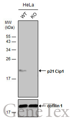
GTX629627 WB Image
Wild-type (WT) and p21 Cip1 knockout (KO) HeLa cell extracts (30 ug) were separated by 15% SDS-PAGE, and the membrane was blotted with p21 Cip1 antibody [GT8611] (GTX629627) diluted at 1:500. The HRP-conjugated anti-mouse IgG antibody (GTX213111-01) was used to detect the primary antibody, and the signal was developed with Trident ECL plus-Enhanced.