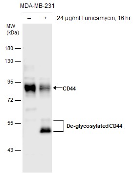
GTX628895 WB Image
Untreated (?) and treated (+) MDA-MB-231 whole cell extracts (30 ug) were separated by 7.5% SDS-PAGE, and the membrane was blotted with CD44 antibody [GT462] (GTX628895) diluted at 1:1000. The HRP-conjugated anti-rabbit IgG antibody (GTX213110-01) was used to detect the primary antibody.
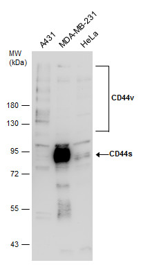
GTX628895 WB Image
Various whole cell extracts (30 ug) were separated by 7.5% SDS-PAGE, and the membrane was blotted with CD44 antibody [GT462] (GTX628895) diluted at 1:1000.
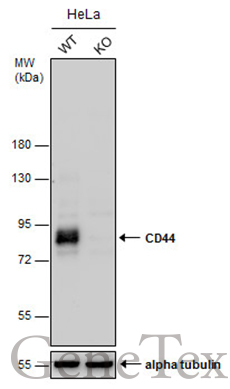
GTX628895 WB Image
Wild-type (WT) and CD44 knockout (KO) HeLa cell extracts (30 ug) were separated by 7.5% SDS-PAGE, and the membrane was blotted with CD44 antibody [GT462] (GTX628895) diluted at 1:500.
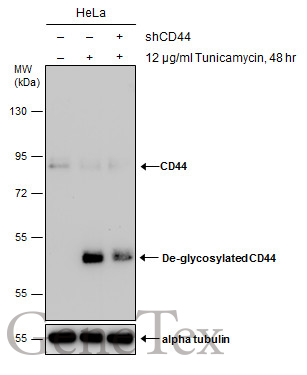
GTX628895 WB Image
Untreated (?) and treated (+) HeLa whole cell extracts (30 ug) were separated by 7.5% SDS-PAGE, and the membrane was blotted with CD44 antibody [GT462] (GTX628895) diluted at 1:1000.
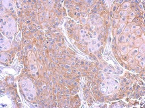
GTX628895 IHC-P Image
CD44 antibody [GT462] detects CD44 protein at membrane on Cal27 xenograft by immunohistochemical analysis.
Sample: Paraffin-embedded Cal27 xenograft.
CD44 antibody [GT462] (GTX628895) dilution: 1:200.
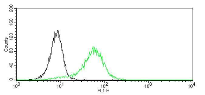
GTX628895 FACS Image
CD44 antibody detects CD44 protein by flow cytometry analysis.
Sample: HL-60 cell fixed in 4% paraformaldehyde at 4oC for 5 min.
Black: Isotype control dilution: 1:100.
Green: CD44 antibody (GTX628895) dilution: 1:100.