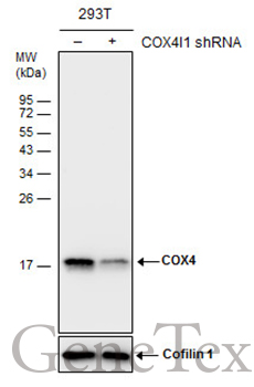
GTX628886 WB Image
Non-transfected (?) and transfected (+) 293T whole cell extracts (30 ug) were separated by 15% SDS-PAGE, and the membrane was blotted with COX4 antibody [GT6310] (GTX628886) diluted at 1:1500.
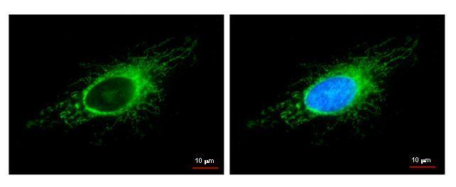
GTX628886 ICC/IF Image
COX4I1 antibody detects COX4I1 protein at mitochondria by immunofluorescent analysis.
Sample: HeLa cells were fixed in -20Åé 100% methanol for 5 min.
Green: COX4I1 protein stained by COX4I1 antibody (GTX628886) diluted at 1:500.
Blue: Hoechst 33342 staining.
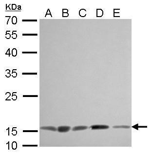
GTX628886 WB Image
COX4 antibody [GT6310] detects COX4I1 protein by Western blot analysis.
A. 30 ug 293T whole cell lysate/extract
B. 30 ug A431 whole cell lysate/extract
C. 30 ug HeLa whole cell lysate/extract
D. 30 ug HepG2 whole cell lysate/extract
E. 30 ug A375 whole cell lysate/extract
12 % SDS-PAGE
COX4 antibody [GT6310] (GTX628886) dilution: 1:1000
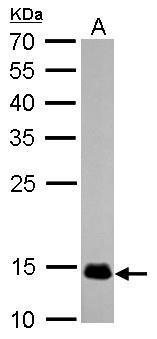
GTX628886 WB Image
COX4 antibody [GT6310] detects COX4I1 protein by Western blot analysis.
A. 50 ug rat muscle lysate/extract
12 % SDS-PAGE
COX4 antibody [GT6310] (GTX628886) dilution: 1:1000
GTX628886 IHC-P Image
COX4 antibody [GT6310] detects COX4I1 protein at cytosol on U87 xenograft by immunohistochemical analysis.
Sample: Paraffin-embedded U87 xenograft.
COX4 antibody [GT6310] (GTX628886) dilution: 1:200.

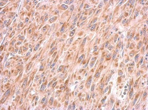
GTX628886 IHC-P Image
COX4 antibody [GT6310] detects COX4I1 protein at cytosol on human hepatoma by immunohistochemical analysis.
Sample: Paraffin-embedded hepatoma.
COX4 antibody [GT6310] (GTX628886) dilution: 1:200.