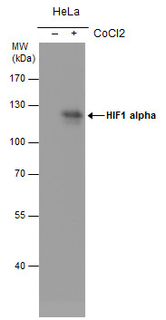
GTX628480 WB Image
HIF1 alpha antibody [GT10211] detects HIF1 alpha protein by western blot analysis. Un-treated (-) and treated (+, 500 uM CoCl2 treatment for 24hr) HeLa whole cell extracts (30 ug) were separated by 7.5% SDS-PAGE, and the membrane was blotted with HIF1 alpha antibody [GT10211] (GTX628480) at a dilution of 1:500. The HRP-conjugated anti-mouse IgG antibody (GTX213111-01) was used to detect the primary antibody.
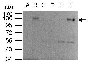
GTX628480 IP Image
HIF-1 alpha antibody immunoprecipitates HIF-1 alpha protein in IP experiments.
IP Sample: 1000 ug HepG2 whole cell lysate/extract
A. 40 ug HepG2 whole cell lysate/extract from normoxia
B. 40 ug HepG2 whole cell lysate/extract from hypoxia 24hr (1%O2)
C. Control with 2.5 ug of preimmune mouse IgG/HepG2 whole cell
lysate/extract from normoxia
D. Control with 2.5 ug of preimmune mouse IgG/ HepG2 whole cell
lysate/extract from hypoxia 24hr (1%O2)
E. Immunoprecipitation of HIF-1 alpha protein by 2.5 ug of HIF-1
alpha antibody (GTX628480)/HepG2 whole cell lysate/extract
from normoxia
F. Immunoprecipitation of HIF-1 alpha protein by 2.5 ug of HIF-1
alpha antibody (GTX628480)/HepG2 whole cell lysate/extract
from hypoxia 24hr (1%O2)
10% SDS-PAGE
The immunoprecipitated HIF-1 alpha protein was detected by HIF-1 alpha antibody (GTX628480) diluted at 1:500.
EasyBlot anti-mouse IgG (GTX221667-01) was used as a secondary reagent.
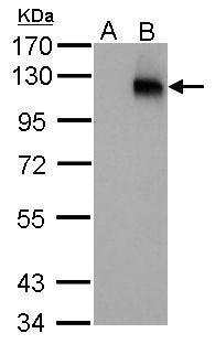
GTX628480 WB Image
HIF1 alpha antibody [GT10211] detects HIF1A protein by western blot analysis.
A. 30 ug HepG2 whole cell lysate/extract (untreated)
B. 30 ug HepG2 whole cell lysate/extract (1%O2 treatment for 48hr)
7.5% SDS-PAGE
HIF1 alpha antibody [GT10211] (GTX628480) dilution: 1:250
The HRP-conjugated anti-mouse IgG antibody (GTX213111-01) was used to detect the primary antibody.
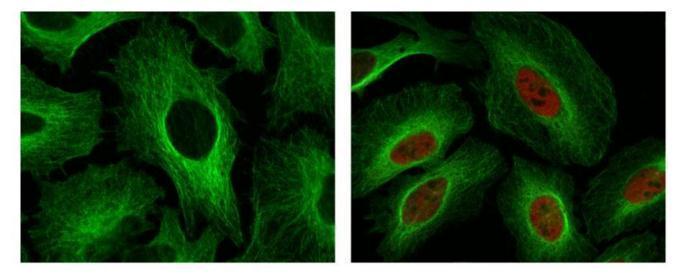
GTX628480 ICC/IF Image
HIF1 alpha antibody detects HIF1 alpha protein at nuclear by confocal immunofluorescent analysis. Sample: Hypoxia (500uM CoCl2) treated 24hr (right) or untreated (left) HeLa cells were fixed in 4% paraformaldehyde for 15 min. Red: HIF1 alpha protein stained by HIF1 alpha antibody (GTX628480) diluted at 1:250. Green: Alpha-tubulin, a cytoskeleton marker, stained by Rabbit Polyclonal antibody (GTX102078) diluted at 1:500. [Images captured by Olympus FV10i Confocal Laser Scanning Microscope]