GTX627420 WB Image
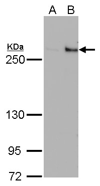
TET1 antibody detects TET1 protein by Western blot analysis.
A. 30 ug 293T whole cell lysate/extract
B. 30 ug whole cell lysate/extract of DDDDK-human TET1-transfected 293T cells
5 % SDS-PAGE
TET1 antibody (GTX627420) dilution: 1:500
GTX627420 WB Image
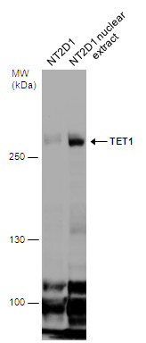
NT2D1 whole cell and nuclear extracts (30 ug) were separated by 5% SDS-PAGE, and the membrane was blotted with TET1 antibody [GT1462] (GTX627420) diluted at 1:500.
GTX627420 WB Image
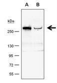
TET1 antibody [GT1462] detects TET1 protein by western blot analysis.
A. 50 ug whole cell lysate/extract from 293T cells transfected with scramble siRNA
B. 50 ug whole cell lysate/extract from TET1-knockdowned 293T cells
6% SDS-PAGE
TET1 antibody [GT1462] (GTX627420) dilution: 1:500
The HRP-conjugated anti-mouse IgG antibody (GTX213111-01) was used to detect the primary antibody.
GTX627420 ICC/IF Image
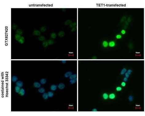
TET1 antibody [GT1462] detects TET1 protein at nucleus by immunofluorescent analysis. Sample: TET1-transfected (right) or untransfected (left) 293T cells were fixed in 4% paraformaldehyde for 15 min. Green: TET1 protein stained by TET1 antibody (GTX627420) diluted at 1:1000. Blue: Hoechst 33342 staining. Scale bar = 10 um.
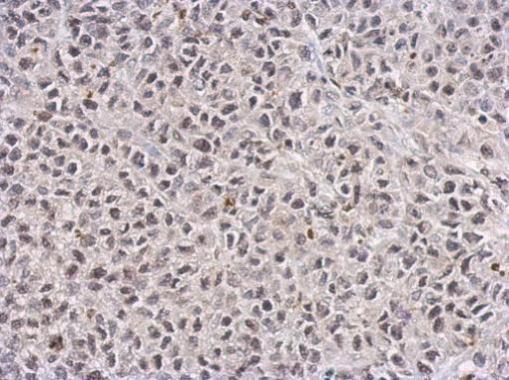
GTX627420 IHC-P Image
TET1 antibody [GT1462] detects TET1 protein at nucleus on HeLa xenograft by immunohistochemical analysis.
Sample: Paraffin-embedded HeLa xenograft.
TET1 antibody [GT1462] (GTX627420) dilution: 1:100.
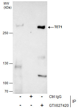
GTX627420 IP Image
Immunoprecipitation of TET1 protein from NT2D1 whole cell extracts using 5 ug of TET1 antibody [GT1462] (GTX627420).
Western blot analysis was performed using TET1 antibody [GT1462] (GTX627420).
EasyBlot anti-Mouse IgG (GTX221667-01) was used as a secondary reagent.