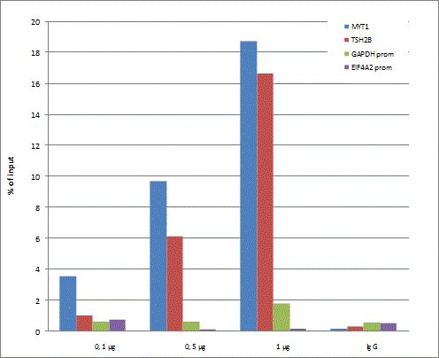
GTX60816 ChIP Image
ChIP was performed with K562 extracts and either control IgG (1 ug) or Histone H3K27me3 (tri-Methy Lys27) antibody (0.1, 0.5, and 1 ug per ChIP experiment). The precipitated DNA was detected by qPCR with primers targeting to positive controls MYT1 and TSH2B genes, and to negative controls GAPDH and EIF4A2 promoters.
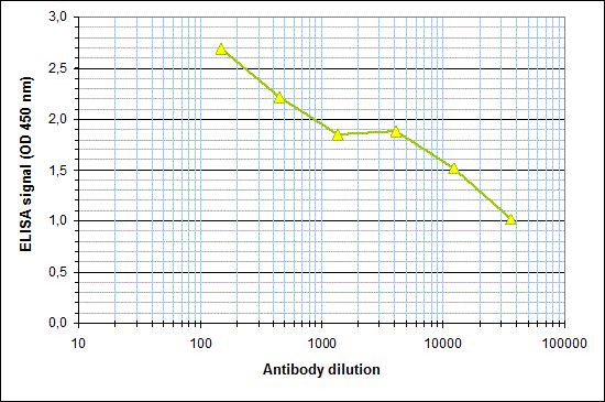
GTX60816 ELISA Image
ELISA was performed using a serial dilution of Histone H3K27me3 (tri-Methy Lys27) antibody in antigen coated wells. By plotting the absorbance against the antibody dilution, the titer of the antibody was estimated to be 1:22,400.

GTX60816 Dot Image
Dot blot analysis of peptides containing modifications or unmodified sequences of histone H3 and H4 using H3K27me3 antibody at a dilution of 1:20,000. One hundred to 0.2 pmol of the respective peptides were spotted on a membrane for analysis.

GTX60816 WB Image
WB analysis of whole cell (25 ug, lane 1) and histone extracts (15 ug, lane 2) from HeLa cells, and 1 ug of recombinant histone H2A, H2B, H3 and H4 (lane 3, 4, 5 and 6, respectively) using H3K27me3 antibody at a dilution of 1:1,000 in TBS-Tween containing 5% skimmed milk.

GTX60816 ICC/IF Image
ICC/IF analysis of NIH3T3 cells using H3K27me3 antibody (green) and DAPI (blue). Cells were fixed with 4% formaldehyde for 10Åf and blocked with PBS/TX-100 containing 5% normal goat serum and 1% BSA. The cells were immunofluorescently labeled with H3K27me3 antibody at a dilution of 1:500.