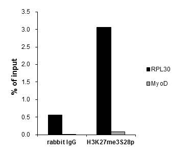
GTX60329 ChIP Image
ChIP was performed with HeLa nuclear extract and either 1 ul of H3K27me3S28p antibody or 5 ug of control rabbit IgG. The precipitated DNA was detected by QPCR with primer set targeting to RPL30 promoter or MyoD.
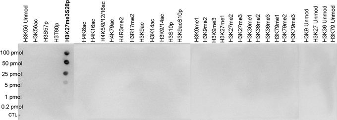
GTX60329 Dot Image
Dot blot analysis of peptides containing modified histone H3 and H4 and unmodified sequences of histone H3 using H3K27me3S28ph antibody at a dilution of 1:20,000. One hundred to 0.2 pmol of the peptides were spotted on a membrane for analysis.
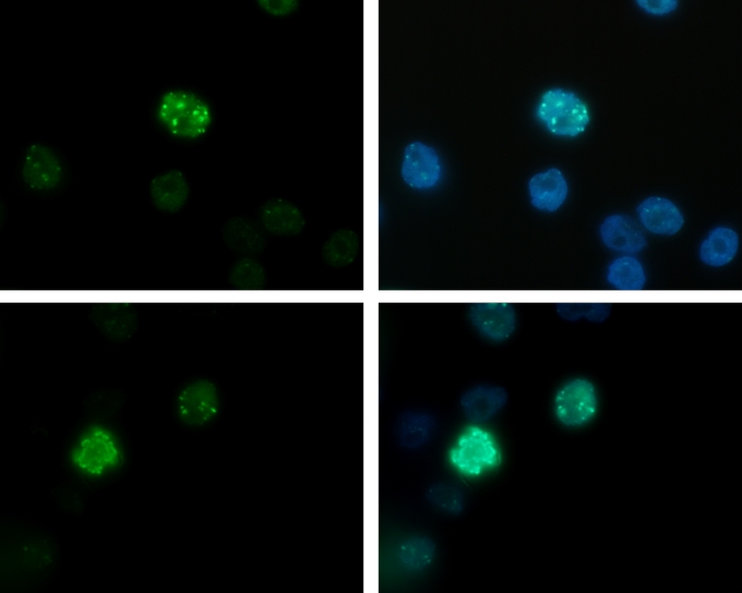
GTX60329 ICC/IF Image
ICC/IF analysis of HeLa asynchronous cells using H3K27me3S28ph antibody and DAPI. Cells were fixed with formaldehyde, permeabilized with sodium citrate and Triton X100 and blocked with PBS containing 2.5% BSA. Cells were immunofluorescently labelled with the H3K27me3S28ph antibody diluted 1:200 (green). The nuclei were stained with DAPI (blue).
GTX60329 IP Image
HeLa cells were treated with colcemid to block the cell cycle in metaphase and were fixed with formaldehyde. Chromatin from 10,000 cells was sheared and used for immunoprecipitation (IP). IP was performed with 5 ul of H3K27me3S28ph antibody. The immunoprecipitated proteins were analysed by WB with the antibody diluted 1:500 in TBS-Tween containing 5% skimmed milk. Lane 1 shows the result of the IP; a negative IP control (no antibody added) and a positive control (sheared chromatin from 10,000 cells) are shown in lane 2 and 3, respectively.
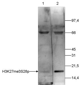
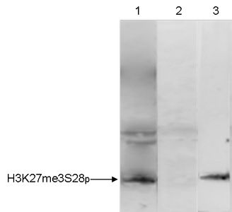
GTX60329 WB Image
WB analysis of histone extracts (15 ug) from HeLa cells using H3K27me3S28ph antibody diluted 1:250 in TBS-Tween containing 5% skimmed milk. Lane 2 shows the result of the WB analysis with the antibody; lane 1 shows the same analysis after incubation of the antibody with 750 pmol blocking peptide for 1 hour at room temperature.
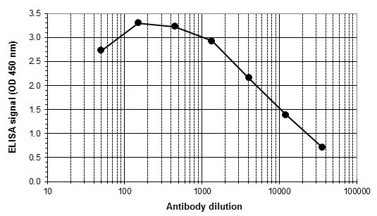
GTX60329 ELISA Image
ELISA was performed using a serial dilution of Histone H3K27me3S28ph (tri-Methyl Lys27, phospho Ser28) antibody in antigen coated wells. By plotting the absorbance against the antibody dilution, the titer of the antibody was estimated to be 1:8,300.